Download
1 / 24
290 likes | 768 Views
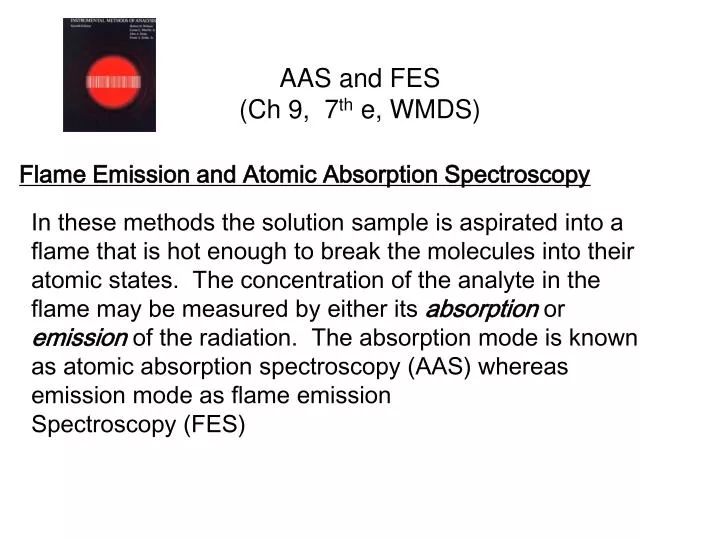
AAS and FES (Ch 9, 7 th e, WMDS). Flame Emission and Atomic Absorption Spectroscopy.

E N D
AAS and FES(Ch 9, 7th e, WMDS) Flame Emission and Atomic Absorption Spectroscopy In these methods the solution sample is aspirated into a flame that is hot enough to break the molecules into their atomic states. The concentration of the analyte in the flame may be measured by either its absorption or emission of the radiation. The absorption mode is known as atomic absorption spectroscopy (AAS) whereas emission mode as flame emission Spectroscopy (FES)
AAS and FES(Ch 9, 7th e, WMDS) Flame Emission and Atomic Absorption Spectroscopy We will look at Atomic Absorption first, though the two methods share many of the same components. Atomic spectroscopy is one of the major tools for analysis of trace (ppm and ppb) metallic elements in industrial and environmental laboratories. There a variety of ways to atomize the analyte. These methods are shown in Table 9.1, page 224.
AAS and FES(Ch 9, 7th e, WMDS) Flame Emission and Atomic Absorption Spectroscopy • One of the features common to both Flame Emission and Atomic Absorption is the use of a flame to atomize the analyte. The flame is generally an acetylene/air or an acetylene/NO2 flame. • The function of the flame differs in the two techniques: • In AAS, the flame serves to convert the analyte into free atoms; the conditions should be such that no ionization occurs.
AAS and FES(Ch 9, 7th e, WMDS) Flame Emission and Atomic Absorption Spectroscopy One of the features common to both Flame Emission and Atomic Absorption is the use of a flame to atomize the analyte. The flame is generally an acetylene/air or an acetylene/N2O flame. 2) In FES the flame also serves to atomize the analyte, but it is also to ionize the atoms so their emission lines are used for identification and quantitation.
AAS and FES(Ch 9, 7th e, WMDS) Upper Diagram shows FES while the lower one shows AAS. In FES the flame also provides the excitation, but in AAS it provides only for the atomization .
AAS and FES(Ch 9, 7th e, WMDS) Instrumentation for an Atomic Absorption Spectrometer. The flowing fuel and air mixture provides the aspiration action drawing the solution sample into the flame.
AAS and FES(Ch 9, 7th e, WMDS) Homework Assignment, due Mon Feb 20 page 255 ff 4, 5, 11
AAS and FES(Ch 9, 7th e, WMDS) Atomic Absorption Spectroscopy In atomic absorption spectroscopy the radiation of the lines of the analyte are produced by the lamp; the presence of the atomic state of the analyte in the flame attenuates or reduces the intensity of the radiation. The absorbance is proportional to the concentration of the analyte, similar to optical spectroscopy.
AAS and FES(Ch 9, 7th e, WMDS) The light source in AAS is the hollow cathode lamp (HCL). The cathode is constructed of the metal or metals of the analysis. Passage of a dc voltage through the lamp produces the specific lines of those elements.
AAS and FES(Ch 9, 7th e, WMDS) Spectra output of the multi-element steel hollow cathode lamp. Note the extremely sharp spectral lines.
AAS and FES(Ch 9, 7th e, WMDS) The bandwidth of the absorption line is much broader than the bandwidth of the spectral lines of the HCL.
AAS and FES(Ch 9, 7th e, WMDS) Details of the atomizer-burner
AAS and FES(Ch 9, 7th e, WMDS) The ultrasonic nebulizer places more sample in the flame and thus lowers the detection limit for most elements by a factor of ten.
AAS and FES(Ch 9, 7th e, WMDS) The sample may also be atomized in AAS using the electrically heated graphite furnace
AAS and FES(Ch 9, 7th e, WMDS) The advantage of the Graphite Furnace for atomization in AAS lies in the smaller sample (microliter range) required and the possibility programming the heating cycle to destroy organic materials before the atomization occurs. In the flame AAS the sample is continuously flowing into the flame and the flame thus dilutes the concentration of atomized atoms, lowering the sensitivity.
AAS and FES(Ch 9, 7th e, WMDS) Although the sensitivity varies according to the element being detected, electrothermal (or the graphite furnace) is generally a 100 times more sensitive. Maximum sensitivity for flame AAS ~1-10 ppb while the maximum sensitivity for the graphite furnace AAS ~ 0.02 – 0.06 ppb. (a ppb = 1 ng/mL or 10-9 g/g)
AAS and FES(Ch 9, 7th e, WMDS) The structure of the flame in laminar flow has cones as shown in Figure 9.5, 231 of the textbook. The temperature and type of the flame (oxidizing or reducing differs in these cones. Thus, the burner may be adjusted vertically so the portion of the flame where the light path can passes may be adjusted. The ratio of fuel to oxidant also affects the type of flame presented to the light path; see Figure 9.6, page 233.
AAS and FES(Ch 9, 7th e, WMDS) The sensitivity depends upon whether the signal can be seen above the background signal (or noise). Generally the height of a signal must exceed the noise by ~ 3 times to be counted as a signal.
AAS and FES(Ch 9, 7th e, WMDS) The matrix effect and the use of standard addition to affect accurate measurements. In the absence of severe matrix effects, a standard calibration curve may be used.
AAS and FES(Ch 9, 7th e, WMDS) 9.6 Interferences with FES and AASThere are 4 types of interferences associated with both FES and AAS:1) Background absorption may be corrected by the use of a continuous light source, generally a deuterium lamp so the effect is made over a wide spectral field and is useful regardless of the specific wavelength for the analysis.
AAS and FES(Ch 9, 7th e, WMDS) 9.6 Interferences with FES and AASThere are 4 types of interferences associated with both FES and AAS:2) Spectral line Interferences arise whenever the line of interest cannot be easily resolved from another element or from a molecular band present in the background. Generally, the bandpass of even very good monochromators is a power of 10 greater than the width of the lines of the HCL. In some cases, amplitude modulation of the HCL helps to detect these interferences.
AAS and FES(Ch 9, 7th e, WMDS) 9.6 Interferences with FES and AASThere are 4 types of interferences associated with both FES and AAS:3) Vaporization Interference arise when some components of the sample solution alters the rate of vaporization. The solution to many of these problems is a hotter flame, such as the use of acetylene/nitrous oxide. Another solution is to pretreat the sample with some other ion that binds more strongly than the analyte. (Example: In the analysis of Ca by AAS, the addition of La or Sr releases the Ca within the flame.)
AAS and FES(Ch 9, 7th e, WMDS) 9.6 Interferences with FES and AASThere are 4 types of interferences associated with both FES and AAS:4) Ionization Effects - At the elevated temperatures that both flames and thermoelectrial units operate, atoms with low ionization potentials are ionized. This ionization lowers the population of atoms available for absorption of light from the HCL. This problem is minimized by suppression, the addition of more easily ionized K, Cs, or Sr to the sample.