Download
1 / 105
2.61k likes | 6.72k Views
Blood collection. Islamic University – Gaza (IUG). Introduction. Hematology : Is defined as the study of blood.

E N D
Blood collection Islamic University – Gaza (IUG)
Introduction Hematology: Is defined as the study of blood. Everybody is familiar with the sight of blood - the red fluid that oozes out of your body when you've sustained a cut or a deep injury, which is slightly denser and approximately 3-4 times more viscous than water.
Blood Volume:- Blood volume is variable, but tend to be about 5- 6 liters , or 7- 8 % of the body weight. Factors such as body size, amount of adipose tissue, and electrolyte concentrations all affect volume. Blood Composition:- Approximately 45% of the blood is composed of formed elements: red blood cells , white blood cells , and platelets. The remaining 55% of the blood is fluid portion, of which approximately 90% is water and 10% is composed of proteins, carbohydrates, vitamins, hormones, enzymes, lipids, and salts
The components of blood can be separated by filtration, however, the most common method of separating blood is to centrifuge (spin) it.Three layers are visible in centrifuged blood. The straw-colored liquid portion, called plasma, forms at the top (~55%). A thin cream-colored layer, called the Buffy coat, forms below the plasma.The Buffy coat consists of white blood cells and platelets. The red blood cells form the heavy bottom portion of the separated mixture (~45%). Note:-
Venipuncture (Phlebotomy) • The process of obtaining intravenous access.
There are three veins most commonly used in venipuncture, or phlebotomy: • The cephalic • The median cubital • The basilic veins • These three veins are found in the antecubital area. • The cephalic vein is found on the lateral, or outside, of the arm. • The median cubital vein,the preferred one to use, is found close to the center. • The basilic vein is located on the inner, or medial part of the antecubital area.
The median cubital vein is the preferred vein for phlebotomy because: • It is usually larger than the other veins. • Best anchored vein (More stationary(. • Veins can move, or roll, which makes it more difficult to perform phlebotomy. The median cubital is typically well anchored, which makes it less likely that the patient will feel pain during phlebotomy, or bruise afterwards.
Median Cubital – First Choice • This vein is located in the antecubital fossa. (the area of the arm in front of the elbow) • Well anchored vein, usually large and prominent. • Very few problems. Offering the best chance for a close to painless puncture, as there are few nerve endings close to this vein.
Cephalic Vein – Second Choice • Cephalic vein which is located on the lateral side of the arm. • This vein is usually well anchored. • The cephalic vein may lie close to the surface.
Basilic Vein – Third Choice • Located on the medial side of the arm. • In many patients this vein may not be well anchored and will roll, making it difficult to access with the needle. • Additionally, this area is often more sensitive, thus a stick is slightly more painful for the patient
The cephalic and basilic veins are only used if the medial cubital vein is not felt. • The cephalic vein is the second choice usually, since it is fairly well anchored. This is often the only vein that can be felt in patients who are obese. • The basilic vein is kept as a last choice option. It rolls more easily and runs directly over a nerve and an artery, making it a more dangerous and painful area to use.
Hand Veins • At times, none of the veins of the antecubital fossa will be felt or not be able to be used due to intravenous placement or injury, hand veins may be used. • Veins of the hand and wrist are usually close to the surface, but they are prone to movement and rolling. • Using these veins tends to be more painful for the patient, since there are nerves running through the hand as well. • If using these veins, it is important to anchor the vein with your hand, holding it in place, when you are drawing the blood.
Venipuncture, why? • Intravenous therapy • Venous blood sample • Parenteral nutrition
Anticoagulants • Most hematology and coagulation procedures must be performed on whole blood or plasma. There for, as soon as the blood is withdrawn from the patient, it is mixed with an anticoagulant to prevent coagulation. The three most commonly used anticoagulants in the hematology laboratory are discussed below: 1- EDTA: • Is generally available as the sodium, dipotassium or tripotassium salt of ethylene diamine tetra acetic acid. It is used in concentration of 1.5(±.25). • EDTA prevents coagulation by binding the calcium in the blood (calcium is required for blood coagulation).
Excessive concentration of EDTA cause: • Shrinkage of the red blood cells leading to decreased hematocrit, increased MCHC, falsely low ESR. • Degenerative changes in the white cells and the platelets will swill and break up causing a falsely increased in platelet counts.
2- Sodium citrate: • Is used for coagulation studies in a concentration of 1 part 0.109M sodium citrate (tri sodium citrate dehydrate) to9 part whole blood. • Sodium citrate prevents coagulation by binding the calcium of the blood in a soluble complex. 3- Heparin: • May be used in concentration of 15 to 30 units/ml of whole blood. its may cause clumping of platelets and white cells. • Coagulation is prevented by interaction with anti thrombin III and subsequent inhibition of thrombin.
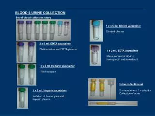
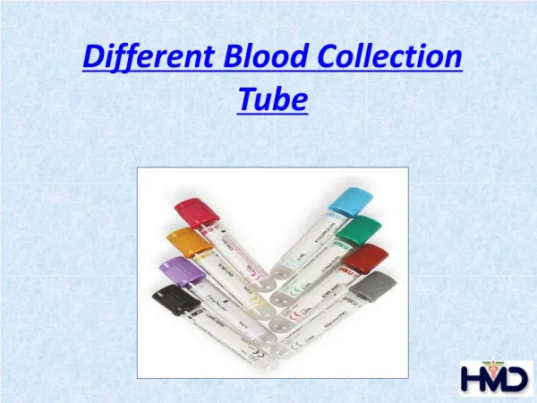
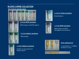
glass or plastic tube with a rubber stopper. It has a vacuum so that blood will flow into the tube. anticoagulants and/or other chemical additives. Blood Collection tubes
Rubber stoppers of blood collection tubes are color coded. Each type of stopper indicates a different additive or a different tube type. Blood collection tubes
The rubber stopper is positioned inside the plastic shield Blood collection tubes: Safety
Needles • Different sizes. • size =gauge. • The larger the needle, the smaller the gauge number. • 21 or 22 gauge needle is mostly used.
Single draw needles are of the type that fit on a syringe, and can be used only to fill the syringe to which they are connected. Single Draw Needle
Winged infusion set Difficult venipunctures including pediatric draws with a syringe or a holder and vacuum collection tube system. 21, 23, or 25 gauge. Butterfly Needle
Lancets are used for difficult venipunctures, including pediatric draws. Lancets
Vein easier to SEE, FEEL and PUNCTURE Tourniquets
Gloves must be worn for all procedures requiring vascular access. Non-powdered latex gloves are most commonly used; Gloves
Greeting • Always greet patient in a professional, friendly manner. • A good initial impression will earn the patients trust, and make it easier and more pleasant to draw a good specimen. • Identify yourself by name and department. • Explain the reason for your presence.
Technical Tip • The more relaxed and trusting your patient, the greater chance of a successful non traumatic venipuncture. • Good verbal, listening, and nonverbal skills are very important for patient reassurance
Patient Identification • Make sure the name, medical record number, and date of birth on your order/requisition match those on the patient’s armband. • Verify the patient’s identity by politely asking them to state their full name. • Properly identifying patients and specimens is probably the single most critical part of your job. • The consequences of misidentifying a specimen can be life threatening. • Never rely on the patient name on the door or above the bed. Patients are frequently moved from room to room.
Technical Tip • A hospitalized patient must always be correctly identified by an ID band that is attached to the patient.
Technical Tip • Patients are often reassured that proper safety measures are being followed when gloves are put on in their presence.
Position the Patient • Comfortable position • Turn the arm so that the wrist and palm face upward, and the antecubital area is accessible
Technical Tip • When supporting the patient’s arm, do not hyperextend the elbow. This may make vein palpation difficult.
Applying the tourniquet • Tie the tourniquet just above the elbow. • The tourniquet should be applied a maximum of 1 – 2 minutes.