Download
1 / 11
110 likes | 330 Views
Cytoskeletal dynamics in vitro. Assembly of actin filaments in vitro The Critical concentration Treadmilling The regulation of actin filament dynamics in vivo. Structures of monomeric G-actin and F-actin. F-actin. G-actin. Actin is the most abundant protein in all eukaryotic cells.

E N D
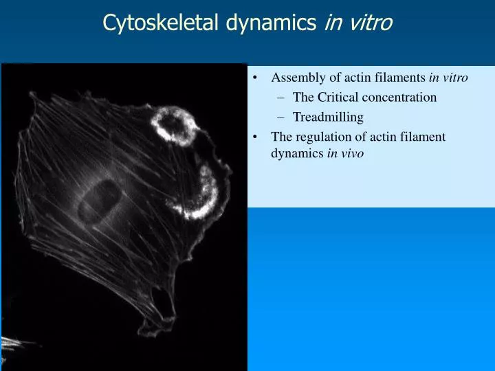
Cytoskeletal dynamics in vitro • Assembly of actin filaments in vitro • The Critical concentration • Treadmilling • The regulation of actin filament dynamics in vivo
Structures of monomeric G-actin and F-actin F-actin G-actin • Actin is the most abundant protein in all eukaryotic cells. • Encoded by a highly conserved gene family • Yeasts and amebas have 1 actin gene; Dictyostelium has 17; Birds and mammals have ~ 6. • Different isoforms exist, e.g. -actin (muscle), -actin (nonmuscle cells) G= globular F= filamentous
Assembled from asymmetric monomers. ATP is required for assembly and is hydrolyzed after subunit addition. K+ and Mg 2+ are required. Assembled actin filaments have polarity, a + end and a - end. This can be detected by myosin head “decoration” experiments. Assembly of actin filaments
Actin filaments have polarity Polarity can be detected by myosin head “decoration” experiments. Actin filament growth is (x10) more rapid at the +end
The dynamics of actin assembly • Actin subunits will polymerize in vitro in the presence of ATP, K+ and Mg 2+ • The degree of polymerization can be monitored by: • Viscometry, sedimentation and fluorescence microscopy. • Polymerization proceeds in 3 phases • 1. Lag phase, where monomers associate into unstable oligomers. • 2. Elongation phase, rapid polymerization of actin monomers onto previously formed oligomers. • 3. Steady - state, polymerization is limited by decreased monomer concentration. An equilibrium is reached between the filaments and actin monomers in solution. The concentration of monomers at which this happens is called the Critical concentration, Cc. • The Critical concentration is a measure of the ability of a solution of G-actin to polymerize. • ATP enhances polymerization at the + end. • Hydrolysis occurs later and promotes disassembly of actin from - ends.
Capping the - end will allow growth only from + end, Cc = 0.1uM Capping the + end will allow growth only from the - end, Cc = 0.8uM Thus when monomer conc. < 0.1uM, no growth occurs. When monomer conc. is between 0.1uM and 0.8uM growth will be only from + end. If mon. conc. > 0.8uM growth occurs at both ends but faster at + end. 0.1uM 0.8 uM The Critical concentration for polymerization is different at + and - ends
Diagram of treadmilling • The length of the filament stays constant but subunits “flux” through the structure from the + to - end.
Treadmilling is the dynamic behavior characteristic of actin filaments • It occurs when the concentration of actin monomers is between the Cc’s of + and - ends. • Monomers add at the + end at the same rate as they are lost at the - end. • ATP-G actin at +end favors growth • ADP-G actin at -end favors disassembly
G-actin F-actin Regulation of actin filament dynamics in vivoFig. 16-52, Alberts • Drugs that stabilize actin filaments, e.g. phalloidins • (bind to sides of f-actin) • Drugs that destabilize actin filaments e.g. cytochalasins • Cytochalasin D (binds +end of f-actin) • Latrunculin (binds G-actin)