Download
1 / 20
200 likes | 366 Views
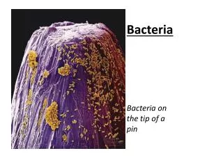
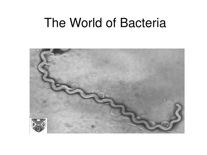
The World of Bacteria. What does a bacterium look like?. Internal Structures: cytoplasm nucleoid ribosomes Boundaries: cell membrane cell wall capsule Appendages: flagellum pili. Shapes of Bacteria. Shapes Bacilli Cocci Sprilli. Growth Patterns

E N D
What does a bacterium look like? Internal Structures: cytoplasm nucleoid ribosomes Boundaries: cell membrane cell wall capsule Appendages: flagellum pili
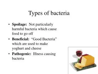
Shapes Bacilli Cocci Sprilli Growth Patterns “Prefixes describe pattern” “strepto…. Means in chains “staphylo…Means in clusters Examples: Streptococcus pneumoniae Bacteria Identification Criteria
The Gram Stain Christian Gram (1884) develops gram staining process. Use of two stains: crystal violet and safranine Stains interact differently with a particular type of cell wall. Cells will be either Gram positive or Gram negative.
How else are bacteria identified? Another criterion? Composition and Construction of the Cell Wall
Gram Negative The Gram-negative cell wall is composed of a thin, inner layer of peptidoglycan and an outer membrane consisting of molecules of phospholipids, lipopolysaccharides (LPS), lipoproteins and sutface proteins. The lipopolysaccharide consists of lipid A and O polysaccharide.
Gram Staining Process What color are gram positive? What color are gram negative?
Which is Gram positive?Which is Gram negative? Gram negative: A group of bacteria that do not retain the crystal violet dye after the differential staining procedure known as Gram staining. They appear pink due to the counterstain, safranin. Gram positive appears purple. The difference between Gram negative and Gram positive bacteria is the cell wall structure, which accounts for the different staining characteristics
The Gram Stain Results: Gram Negative Gram Positive So what is the medical significance of this technique?
What are antibiotics? Antibiotics are strong medicines. Antibiotics only work against infections caused by bacteria. Antibiotics kill bacteria or stop them from growing. Antibiotics should be used wisely.
Antibiotic Sensitivity Figure F. Antibiotic-sensitivity testing. Petri dishes were spread-inoculated with Staphylococcus albus (white growth) or Micrococcus luteus (yellow growth) before antibiotic assay "rings" were placed on the agar surface. The coloured disks at the end of each spoke of the rungs are impregnated with different antibiotics. Clockwise from the top (arrow) these are: Novobiocin, Penicillin G, Streptomycin (white disk), Tetracycline, Chloramphenicol, Erythromycin, Fusidic acid (green disk) and Methicillin. Clear zones of suppression of bacterial growth around the individual antibiotic disks are evidence of sensitivity to these antibiotics. The diameter of the clear zone is related to the initial antibiotic concentration (which differs for the antibiotics on the ring), its solubility and its diffusion rate through agar. Standard tests performed on many bacteria by the manufacturers of these assay disks enable the diameter of the clearing zone to be related to the minimum inhibitory concentration (MIC) of each antibiotic for the strain being tested. The MIC can then be compared with the known tissue levels of these antibiotics when they are administered to patients, to assess whether the antibiotics would be effective for treatment of particular pathogens.
Antibiotic Resistant Bacteria Bacteria that is not affected by an antibiotic. Antibiotic resistance is a phenotype. They posses a gene that renders them resistant (genotype). Antibiotic resistant gene produces enzymes that breakdown the antibiotics.
How Antibiotic Resistant Bacteria Develop http://www.fda.gov/fdac/features/795_antibio.html Antibiotic resistance results from gene action. Bacteria acquire genes conferring resistance in any of three ways. In spontaneous DNA mutation, bacterial DNA (genetic material) may mutate (change) spontaneously (indicated by starburst). Drug-resistant tuberculosis arises this way. In a form of microbial sex called transformation, one bacterium may take up DNA from another bacterium. Penicillin-resistant gonorrhea results from transformation
One More Way towards Resistance Most frightening, however, is resistance acquired from a small circle of DNA called a plasmid, that can flit from one type of bacterium to another. A single plasmid can provide a slew of different resistances. In 1968, 12,500 people in Guatemala died in an epidemic of Shigella diarrhea. The microbe harbored a plasmid carrying resistances to four antibiotics! What on the plasmid makes the bacteria resistant?
Resistant Genes Genes are located in a circular piece of DNA found in the bacterial cell called a plasmid. Bacterial also has chromosomal DNA.