Download
1 / 124
1.26k likes | 1.52k Views
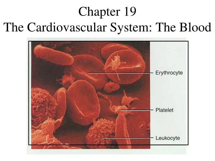
Chapter 19 The Cardiovascular System: The Blood. Fluids of the Body. Cells of the body are serviced by 2 fluids blood composed of plasma and a variety of cells transports nutrients and wastes interstitial fluid bathes the cells of the body

E N D
Fluids of the Body • Cells of the body are serviced by 2 fluids • blood • composed of plasma and a variety of cells • transports nutrients and wastes • interstitial fluid • bathes the cells of the body • Nutrients and oxygen diffuse from the blood into the interstitial fluid & then into the cells • Wastes move in the reverse direction • Hematology is study of blood and blood disorders
Functions of Blood • Transportation • O2, CO2, metabolic wastes, nutrients, heat & hormones • Regulation • helps regulate pH through buffers • helps regulate body temperature • coolant properties of water • vasodilatation of surface vessels dump heat • helps regulate water content of cells by interactions with dissolved ions and proteins • Protection from disease & loss of blood
Physical Characteristics of Blood • Thicker (more viscous) than water and flows more slowly than water • Temperature of 100.4 degrees F • pH 7.4 (7.35-7.45) • 8 % of total body weight • Blood volume • 5 to 6 liters in average male • 4 to 5 liters in average female • hormonal negative feedback systems maintain constant blood volume and osmotic pressure
Components of Blood • Hematocrit • 55% plasma • 45% cells • 99% RBCs • < 1% WBCs and platelets
Blood Plasma • 0ver 90% water • 7% plasma proteins • created in liver • confined to bloodstream • albumin • maintain blood osmotic pressure • globulins (immunoglobulins) • antibodies bind to foreignsubstances called antigens • form antigen-antibody complexes • fibrinogen • for clotting • 2% other substances • electrolytes, nutrients, hormones, gases, waste products
Formed Elements of Blood • Red blood cells ( erythrocytes ) • White blood cells ( leukocytes ) • granular leukocytes • neutrophils • eosinophils • basophils • agranular leukocytes • lymphocytes = T cells, B cells, and natural killer cells • monocytes • Platelets (special cell fragments)
Hematocrit • Percentage of blood occupied by cells • female normal range • 38 - 46% (average of 42%) • male normal range • 40 - 54% (average of 46%) • testosterone • Anemia • not enough RBCs or not enough hemoglobin • Polycythemia • too many RBCs (over 65%) • dehydration, tissue hypoxia, blood doping in athletes
Red Blood Cells or Erythrocytes • Contain oxygen-carrying protein hemoglobin that gives blood its red color • 1/3 of cell’s weight is hemoglobin • Biconcave disk 8 microns in diameter • increased surface area/volume ratio • flexible shape for narrow passages • no nucleus or other organelles • no cell division or mitochondrial ATP formation • Normal RBC count • male 5.4 million/drop ---- female 4.8 million/drop • new RBCs enter circulation at 2 million/second
Hemoglobin • Globin protein consisting of 4 polypeptide chains • One heme pigment attached to each polypeptide chain • each heme contains an iron ion (Fe+2) that can combine reversibly with one oxygen molecule
RBC Life Cycle • RBCs live only 120 days • wear out from bending to fit through capillaries • no repair possible due to lack of organelles • Worn out cells removed by fixed macrophages in spleen & liver • Breakdown products are recycled
Erythropoiesis: Production of RBCs • Proerythroblast starts to produce hemoglobin • Many steps later, nucleus is ejected & a reticulocyte is formed • orange in color with traces of visible rough ER • Reticulocytes escape from bone marrow into the blood • In 1-2 days, they eject the remaining organelles to become a mature RBC
WBC Anatomy and Types • All WBCs (leukocytes) have a nucleus and no hemoglobin • Granular or agranular classification based on presence of cytoplasmic granules made visible by staining • granulocytes are neutrophils, eosinophils or basophils • agranulocytes are monocyes or lymphocytes
WBC Physiology • Less numerous than RBCs • 5000 to 10,000 cells per drop of blood • 1 WBC for every 700 RBC • Leukocytosis is a high white blood cell count • microbes, strenuous exercise, anesthesia or surgery • Leukopenia is low white blood cell count • radiation, shock or chemotherapy • Only 2% of total WBC population is in circulating blood at any given time • rest is in lymphatic fluid, skin, lungs, lymph nodes & spleen
Differential WBC Count • Detection of changes in numbers of circulating WBCs (percentages of each type) • indicates infection, poisoning, leukemia, chemotherapy, parasites or allergy reaction • Normal WBC counts • neutrophils 60-70% (up if bacterial infection) • lymphocyte 20-25% (up if viral infection) • monocytes 3 -- 8 % (up if fungal/viral infection) • eosinophil 2 -- 4 % (up if parasite or allergy reaction) • basophil <1% (up if allergy reaction or hypothyroid)
Bone Marrow Transplant • Intravenous transfer of healthy bone marrow • Procedure • destroy sick bone marrow with radiation & chemotherapy • donor matches surface antigens on WBC • put sample of donor marrow into patient's vein for reseeding of bone marrow • success depends on histocompatibility of donor & recipient • Treatment for leukemia, sickle-cell, breast, ovarian or testicular cancer, lymphoma or aplastic anemia
Platelets--Life History • Platelets form in bone marrow by following steps: • myeloid stem cells to megakaryocyte-colony forming cells to megakaryoblast to megakaryocytes whose cell fragments form platelets • Short life span (5 to 9 days in bloodstream) • formed in bone marrow • few days in circulating blood • aged ones removed by fixed macrophages in liver and spleen
Complete Blood Count • Screens for anemia and infection • Total RBC, WBC & platelet counts; differential WBC; hematocrit and hemoglobin measurements • Normal hemoglobin range • infants have 14 to 20 g/100mL of blood • adult females have 12 to 16 g/100mL of blood • adult males have 13.5 to 18g/100mL of blood
Hemostasis • Stoppage of bleeding in a quick & localized fashion when blood vessels are damaged • Prevents hemorrhage (loss of a large amount of blood) • Methods utilized • vascular spasm • platelet plug formation • blood clotting (coagulation = formation of fibrin threads)
Blood Clotting • Blood drawn from the body thickens into a gel • gel separates into liquid (serum) and a clot of insoluble fibers (fibrin) in which the cells are trapped • If clotting occurs in an unbroken vessel is called a thrombosis • Substances required for clotting are Ca+2, enzymes synthesized by liver cells and substances released by platelets or damaged tissues • Clotting is a cascade of reactions in which each clotting factor activates the next in a fixed sequence resulting in the formation of fibrin threads • prothrombinase & Ca+2 convert prothrombin into thrombin • thrombin converts fibrinogen into fibrin threads
Overview of the Clotting Cascade • Prothrombinase is formed by either the intrinsic or extrinsic pathway • Final common pathway produces fibrin threads
Clot Retraction & Blood Vessel Repair • Clot plugs ruptured area of blood vessel • Platelets pull on fibrin threads causing clot retraction • trapped platelets release factor XIII stabilizing the fibrin threads • Edges of damaged vessel are pulled together • Fibroblasts & endothelial cells repair the blood vessel
Anemia = Not Enough RBCs • Symptoms • oxygen-carrying capacity of blood is reduced • fatigue, cold intolerance & paleness • lack of O2 for ATP & heat production • Types of anemia • iron-deficiency =lack of absorption or loss of iron • pernicious = lack of intrinsic factor for B12 absorption • hemorrhagic = loss of RBCs due to bleeding (ulcer) • hemolytic = defects in cell membranes cause rupture • thalassemia = hereditary deficiency of hemoglobin • aplastic = destruction of bone marrow (radiation/toxins)
Sickle-cell Anemia (SCA) • Genetic defect in hemoglobin molecule (Hb-S) that changes 2 amino acids • at low very O2 levels, RBC is deformed by changes in hemoglobin molecule within the RBC • sickle-shaped cells rupture easily = causing anemia & clots • Found among populations in malaria belt • Mediterranean Europe, sub-Saharan Africa & Asia • Person with only one sickle cell gene • increased resistance to malaria because RBC membranes leak K+ & lowered levels of K+ kill the parasite infecting the red blood cells
Hemophilia • Inherited deficiency of clotting factors • bleeding spontaneously or after minor trauma • subcutaneous & intramuscular hemorrhaging • nosebleeds, blood in urine, articular bleeding & pain • Hemophilia A lacks factor VIII (males only) • most common • Hemophilia B lacks factor IX (males only) • Hemophilia C (males & females) • less severe because alternate clotting activator exists • Treatment is transfusions of fresh plasma or concentrates of the missing clotting factor
Leukemia • Acute leukemia • uncontrolled production of immature leukocytes • crowding out of normal red bone marrow cells by production of immature WBC • prevents production of RBC & platelets • Chronic leukemia • accumulation of mature WBC in bloodstream because they do not die • classified by type of WBC that is predominant---monocytic, lymphocytic.
Chapter 20The Cardiovascular System: The Heart • Heart pumps over 1 million gallons per year • Over 60,000 miles of blood vessels
Heart Location Anterior surface of heart • Heart is located in the mediastinum • area from the sternum to the vertebral column and between the lungs
Heart Orientation • Apex - directed anteriorly, inferiorly and to the left • Base - directed posteriorly, superiorly and to the right • Anterior surface - deep to the sternum and ribs • Inferior surface - rests on the diaphragm • Right border - faces right lung • Left border (pulmonary border) - faces left lung
Heart Orientation • Heart has 2 surfaces: anterior and inferior, and 2 borders: right and left
Surface Projection of the Heart • Superior right point at the superior border of the 3rd right costal cartilage • Superior left point at the inferior border of the 2nd left costal cartilage 3cm to the left of midline • Inferior left point at the 5th intercostal space, 9 cm from the midline • Inferior right point at superior border of the 6th right costal cartilage, 3 cm from the midline
Pericardium • Fibrous pericardium • dense irregular CT • protects and anchors the heart, prevents overstretching • Serous pericardium • thin delicate membrane • contains • parietal layer-outer layer • pericardial cavity with pericardial fluid • visceral layer (epicardium)
Layers of Heart Wall • Epicardium • visceral layer of serous pericardium • Myocardium • cardiac muscle layer is the bulk of the heart • Endocardium • chamber lining & valves
Chambers and Sulci of the Heart • Four chambers • 2 upper atria • 2 lower ventricles • Sulci - grooves on surface of heart containing coronary blood vessels and fat • coronary sulcus • encircles heart and marks the boundary between the atria and the ventricles • anterior interventricular sulcus • marks the boundary between the ventricles anteriorly • posterior interventricular sulcus • marks the boundary between the ventricles posteriorly
Chambers and Sulci Anterior View
Chambers and Sulci Posterior View
Right Atrium • Receives blood from 3 sources • superior vena cava, inferior vena cava and coronary sinus • Interatrial septum partitions the atria • Fossa ovalis is a remnant of the fetal foramen ovale • Tricuspid valve • Blood flows through into right ventricle • has three cusps composed of dense CT covered by endocardium
Right Ventricle • Forms most of anterior surface of heart • Papillary muscles are cone shaped trabeculae carneae (raised bundles of cardiac muscle) • Chordae tendineae: cords between valve cusps and papillary muscles • Interventricular septum: partitions ventricles • Pulmonary semilunar valve: blood flows into pulmonary trunk
Left Atrium • Forms most of the base of the heart • Receives blood from lungs - 4 pulmonary veins (2 right + 2 left) • Bicuspid valve: blood passes through into left ventricle • has two cusps • to remember names of this valve, try the pneumonic LAMB • Left Atrioventricular, Mitral, or Bicuspid valve
Left Ventricle • Forms the apex of heart • Chordae tendineae anchor bicuspid valve to papillary muscles (also has trabeculae carneae like right ventricle) • Aortic semilunar valve: • blood passes through valve into the ascending aorta • just above valve are the openings to the coronary arteries
Myocardial Thickness and Function • Thickness of myocardium varies according to the function of the chamber • Atria are thin walled, deliver blood to adjacent ventricles • Ventricle walls are much thicker and stronger • right ventricle supplies blood to the lungs (little flow resistance) • left ventricle wall is the thickest to supply systemic circulation
Thickness of Cardiac Walls Myocardium of left ventricle is much thicker than the right.
Atrioventricular Valves Open • A-V valves open and allow blood to flow from atria into ventricles when ventricular pressure is lower than atrial pressure • occurs when ventricles are relaxed, chordae tendineae are slack and papillary muscles are relaxed
Atrioventricular Valves Close • A-V valves close preventing backflow of blood into atria • occurs when ventricles contract, pushing valve cusps closed, chordae tendinae are pulled taut and papillary muscles contract to pull cords and prevent cusps from everting
Semilunar Valves • SL valves open with ventricular contraction • allow blood to flow into pulmonary trunk and aorta • SL valves close with ventricular relaxation • prevents blood from returning to ventricles, blood fills valve cusps, tightly closing the SL valves
Valve Function Review Which side is anterior surface? What are the ventricles doing?
Valve Function Review Ventricles contract, blood pumped into aorta and pulmonary trunk through SL valves Atria contract, blood fills ventricles through A-V valves
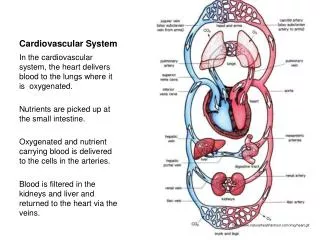
Blood Circulation • Two closed circuits, the systemic and pulmonic • Systemic circulation • left side of heart pumps blood through body • left ventricle pumps oxygenated blood into aorta • aorta branches into many arteries that travel to organs • arteries branch into many arterioles in tissue • arterioles branch into thin-walled capillaries for exchange of gases and nutrients • deoxygenated blood begins its return in venules • venules merge into veins and return to right atrium
Blood Circulation (cont.) • Pulmonary circulation • right side of heart pumps deoxygenated blood to lungs • right ventricle pumps blood to pulmonary trunk • pulmonary trunk branches into pulmonary arteries • pulmonary arteries carry blood to lungs for exchange of gases • oxygenated blood returns to heart in pulmonary veins
Blood Circulation • Blood flow • blue = deoxygenated • red = oxygenated