Download
1 / 71
710 likes | 795 Views
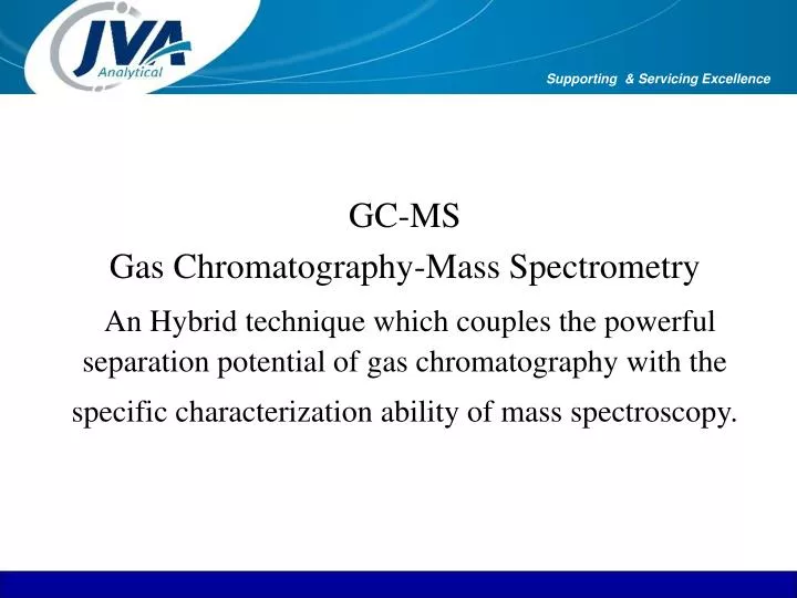
GC-MS Gas Chromatography-Mass Spectrometry An Hybrid technique which couples the powerful separation potential of gas chromatography with the specific characterization ability of mass spectroscopy. Supporting & Servicing Excellence. Overview. GC History What is GC Key Components

E N D
GC-MSGas Chromatography-Mass SpectrometryAn Hybrid technique which couples the powerful separation potential of gas chromatography with the specific characterization ability of mass spectroscopy. Supporting & Servicing Excellence
Overview • GC History • What is GC • Key Components • Separation Process • GC Theory • Carrier Gas • Injectors • Columns
GC History • Development of GC (1941) by Martin and Synge • Theory of Capillary GC (1957) by Golay • Capillary GC Instruments (1977) • Fused Silica Capillary Columns (1979)
What is GC? • GC is a Separation Technique • Sample is usually a complex mixture we require to separate into constituent components. • Why: usually to quantify some or all components e.g. Pharmaceuticals, Environmental pollutants, etc • Occasionally as a qualitative tool
What is the sample? • Usually a mixture of several components • Sample usually introduced as a liquid • Components of interest (analytes) usually in low concentrations (<1% to ppb levels) • Samples dissolved in volatile solvent
Comaparison: GC & HPLC HPLC • non-volatile samples • thermally unstable compounds • macromolecules • inorganic and ionic samples • More complex interface to Mass Spec . • GC • volatile & thermally stable • rapid analysis • good resolution • easily interfaced to Mass Spec
Key components of GC • Hardware to introduce the sample • Technique to separate the sample into components • Hardware to detect the individual components. • Data Processing to process this information.
Separation Process • Sample is introduced into system via hot, vaporising injector. • Typically 1ul injected • Flow of “Carrier Gas” moves vaporised sample (i.e. gas) onto column • Column is coated with wax type material with varying affinity for components of interest • Components are separated in the column based on this affinity. • Individual analytes are detected as they emerge from the end of the column through the Detector.
Example Chromatogram (Capillary) Detector Response Inject Point Time
Analysis of Halogenated Pesticides 4 10 7 11 6 8 12 13 9 14 1 5 15 17 18 3 16 2 2ppb in Water
GC Step by Step • Carrier Gas • Injector • Column • Capillary • Stationary Phase • Detectors • Mass Spectrometer
Carrier Gas • Inert • Helium • Choice dictated by detector, cost, availability • Pressure regulated for constant inlet pressure • Flow controlled for constant flow rate • Chromatographic grade gases (high purity)
Column Types Capillary Columns Length: 10m to 100m Diameter: 180um, 250um, 320um & 530um I.d Packed Columns Length: <2m Diameter: 1/8” & ¼” OD
Typical column flow rates • Capillary Column Flow • 250 um 1 ml/min • 320 um 1.5 ml/min • 530um up to 2.0 ml/min
Purpose of Injection • Deposit the sample into the column in the narrowest band possible • The shorter the band at the beginning of the chromatographic process - tall narrow peaks • Gives maximum resolution and sensitivity • Therefore type of injection method and operating conditions is critical in obtaining precise and accurate results
Splitless injector Design • Graphite/Viton Seal • Reduced Sample Contact • “Unique”Dual Split Vent design • Improved Precision • More Efficient Sweep • Large Internal Volume • Minimum Solvent Tailing • Shortened Capillary Guide • Minimal Cold Spots • Minimal Upswept Volume
Cross Section of PTV Injector Modern Temperature Programmable Injector (Varian 1079) Programmable Temperature Vapourising Injector
Split & Splitless Injection • Most common method of Injection into Capillary Columns • Most commonly misunderstood also! • Same injector hardware is used for both techniques • Electronically controlled Solenoid changes Gas Flow to determine Injector function.
Split Injection • Mechanism by which a portion of the injected solution is discarded. • Only a small portion (1/1000 - 1/20) of sample goes through the column • Used for concentrated samples (>0.1%) • Can be performed isothermally • Fast injection speed • Injector and septa contamination not usually noticed
Splitless Injection • Most of the sample goes through the column (85-100%) • Used for dilute samples (<0.1%) • Injection speed slow • Should not be performed isothermally • Solvent focusing is important • Controlled by solenoid valve • Requires careful optimisation
On Column Injection • All of the sample is transferred to the column • Needle is inserted directly into column or into insert directly above column • Trace analysis • Thermally labile compounds e.g Pesticides, Drugs • Wide boiling point range • High molecular weight
Large Volume Injection • To enhance sensitivity in Envoirnmental applications. • Uses 100µL syringe: Inject up to 70 µl • Very slow injection with injector temperature a few degrees below solvent boiling point, split open, flow at about 150 mls/ min • Solvent vents out of split vent, thus concentrating the analytes • Close split • Fast temperature ramp to top column temperature +20°C • Column programming as per sample requirements
Material of Construction • Metal (1957) • Glass (1959) • Fused Silica (1979) • Aluminium Clad (1984) • Inert Metal (1990)
Capillary Column Characteristics • Length (10M - 50M) • Internal Diameter (0.1mm - 0.53mm) • Liquid Stationary Phase • Film Thickness (0.1um - 5um) • Polarity (Non-polar - Polar)
Stationary Phases • Choice of phase determines selectivity • Hundred of phases available • Many phases give same separation • Same phase may have multiple brand names • Stationary phase selection for capillary columns much simpler • Like dissolves like • Use polar phases for polar components • Use non-polar phases for non-polar components
Column Bleed • Bleed increases with film thickness • Polar columns have higher bleed • Bleed is excessive when column is damaged or degraded • Avoid strong acids or bases • Adhere to manufacturer’s recommended temperature limits • Avoid leaks
Choosing a Column • Internal Diameter • Film Thickness • Length • Phase
Internal Diameter, Smaller ID’s • Good resolution of early eluting compounds • Longer analysis times • Limited dynamic range
ID Effects - larger ID’s • Have less resolution of early eluting compounds • Shorter analysis times • Sufficient resolution for complex mixtures • Greater dynamic range
Film Thickness • Amount of stationary phase coating • Affects retention and capacity • Thicker films increase retention and capacity • Thin films are useful for high boilers • Standard capillary columns typically 0.25µm • 0.53mm ID (Megabore) typically 1.0 - 1.5µm
Column Capacity • The maximum amount that can be injected without significant peak distortion • Column capacity increases with :- • film thickness • temperature • internal diameter • stationary phase selectivity • If exceeded, results in :- • peak broadening • asymmetry • leading
Length effects - isothermal analysis • Retention more dependant on length • Doubling column length doubles analysis times • Resolution a function of Square Root of Length • Gain 41% in resolution • Is it worth the extra time and expense?-
Length effects - programmed analysis • Retention more dependant on temperature • Marginally increases analysis times • Run conditions should be optimised
Overview • Basic Mass Spectrometry Theory • Types of Ionisation • - Electronic Ionisation • - Chemical Ionisation • Interpretation of Mass Spectra • Ion Trap Theory • Components of the Ion Trap
Basic Mass Spec.Theory • Mass Spec. is a Microanalytical Technique used to obtain information regarding structure and Molecular weight of an analyte • Destructive method ie sample consumed during analysis • In all cases some form of energy is transferred to analyte to cause ionisation • In principle each Mass Spectrum is unique and can be used as a “fingerprint” to characterise the sample • GC/MS is a combination technique that combines the separation ability of the GC with the Detection qualities of Mass Spec.
Basic GCMS Theory(1) • Sample injected onto column via injector • GC then separates sample molecules • Effluent from GC passes through transfer line into the Ion Trap/Ion source • Molecules then undergo electron /chemical ionisation • Ions are then analysed according to their mass to charge ratio • Ions are detected by electron multiplier which produces a signal proportional to ions detected
Basic GCMS Theory(2) • Electron multiplier passes the ion current signal to system electronics • Signal is amplified • Result is digitised • Results can be further processed and displayed
Types of Ionisation • Electron impact ionisation • Chemical Ionisation
Electron Ionisation(1) • Sample of interest vaporised into mass spec • Energy sufficient for Ionisation and Fragmentation of analyte molecules is acquired by interaction with electrons from a hot Filament • 70 eV is commonly used • Source of electrons is a thin Rhenium wire heated electrically to a temp where it emits free electrons
Electron Ionisation • The physics behind mass spectrometry is that a charged particle passing through a magnetic field is deflected along a circular path on a radius that is proportional to the mass to charge ratio, m/e. In an electron impact mass spectrometer, a high energy beam of electrons is used to displace an electron from the organic molecule to form a radical cation known as the molecular ion. If the molecular ion is too unstable then it can fragment to give other smaller ions. The collection of ions is then focused into a beam and accelerated into the magnetic field and deflected along circular paths according to the masses of the ions. By adjusting the magnetic field, the ions can be focused on the detector and recorded.
Chemical ionisation • Used to confirm molecular weight • Known as a “soft” ionisation technique • Differs from EI in that molecules are ionised by interaction or collision with ions of a reagent gas rather that with electrons • Common reagent gases used are Methane , Isobutane and Ammonia • Reagent gas is pumped directly into ionisation chamber and electrons from Filament ionise the reagent gas
Chemical Ionisation(2) • First - electron ionization of CH4: • CH4 + e- CH4+ + 2e- • Fragmentation forms CH3+, CH2+, CH+ • Second - ion-molecule reactions create stable reagent ions: • CH4+ + CH4 CH3 + CH5+ • CH3+ + CH4 H2 + C2H5+ • CH5+ and C2H5+ are the dominant methane CI reagent ions