Download
1 / 56
600 likes | 938 Views
Acute kidney Injury(AKI) . Dr Dana Ahmed Sharif Renal Physician MRCP UK/ MRCP London. Resource materials. Davidson’s Principles & Practice of Medicine Kumar & Clark Clinical Medicine Oxford textbook of Clinical Nephrology Oxford handbook of Nephrology and hypertension

E N D
Acute kidney Injury(AKI) Dr Dana Ahmed Sharif Renal Physician MRCP UK/ MRCP London
Resource materials • Davidson’s Principles & Practice of Medicine • Kumar & Clark Clinical Medicine • Oxford textbook of Clinical Nephrology • Oxford handbook of Nephrology and hypertension • Renal Association website( www.renal.org) • K/DOQI guideline (www.kidney.org) • AKI network ( www.akinet.org)
Renal function • Kidney has many roles:
Renal function • Kidney has many roles: - Excretory function
Renal function • Kidney has many roles: - Excretory function - Osmolality regulation
Renal function • Kidney has many roles: - Excretory function - Osmolality regulation - Acid base balance
Renal function • Kidney has many roles: - Excretory function - Osmolality regulation - Acid base balance - BP regulation through salt and water balance
Renal function • Kidney has many roles: - Excretory function - Osmolality regulation - Acid base balance - BP regulation through Salt and water balance - Hormone secretion ( Erythropoietin, Vit D3)
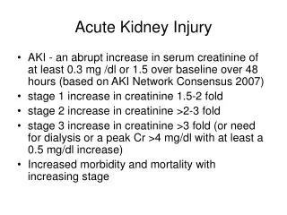
Definition of Acute Kidney Injury Acute usually reversible decline in renal function* • Rapid time course( < 48 hrs) • Reduction of kidney function: A- Rise in serum creatinine, defined by either: 1- absolute increase in serum creatinine of >0.3mg/dl( >26µmol/l) 2- % increase in serum creatinine of > 50% B- Reduction in urine output, defined as < 0.5ml/kg/hr for more than 6 hrs * Acute kidney injury network
Incidence of AKI* • 500 ppm/year – UK ( up to 38,000/yr) • Incidence of AKI needing dialysis 200 ppm/year • Pre renal and acute tubular necrosis (ATN) accounts for 75% of the cases of AKI • 7% of all hospital admissions( 65% of intensive care admission) • Mortality: • 5-10% in uncomplicated AKI • 50-70% in AKI secondary to other organ failure( intensive care) • > 50% in dialysis requiring AKI *XueJL, Daniels F, Star RA et al. Incidence and mortality of acute renal failure in Medicare beneficiaries, 1992 to 2001. J Am Soc Nephrol 2006; 17: 1135–1142.
Diagnosing pre-renal AKI • Is the patient volume depleted?
Diagnosing pre-renal AKI • Is the patient volume depleted? • Is cardiac function good?
Diagnosing pre-renal AKI • Is the patient volume depleted? • Is cardiac function good? • Is the patient septic?
Diagnosing pre-renal AKI • Is the patient volume depleted? • Is cardiac function good? • Is the patient septic? History Examination Investigation
Diagnosing pre renal AKI • History • Examination : 1- Signs of Hypovolaemia: a- Low BP( and reduced pulse pressure) b- Postural BP drop ( a fall in systolic BP > 10mmHg) c- Sinus tachycardia and postural increase in heart rate ( increase in HR > 10 beat/min). d- Low JVP even when the patient is supine e- Cool peripheries and vasoconstriction f- Poor urine output
Diagnosing pre-renal AKI 2- Sings of hypervolaemia( high extracellular fluid): a- Increased circulating volume: - High BP - Elevation of the JVP b- Increased interstitial fluid: - Peripheral or generalized oedema - Pulmonary oedema (tachypnoea, tachycardia, third heart sound, basal crackles) - Pleural effusion - Ascites . Lab investigation: - Blood tests - urine: including urinary Na( low)
Case 1 • 67 yr man – IHD • Admitted with D&V – O/E JVP not seen, BP 100/60 lying, 80/50 standing, pulse 105 bpm • Creatinine 5.8 (0.7-1.2mg/dl) • x2 IV access • Given IV saline • Catheterised and started on furosemide • Function worsened and transferred to renal unit
What was the only helpful intervention 1- Inserting a urinary catheter 2- Inserting a CVP line 3- Administering IV fluids 4- Administering diuretics
What was the only helpful intervention 1- Inserting a urinary catheter 2- Inserting a CVP line 3- Administering IV fluids 4- Administering diuretics
Treatment of pre renal failure • DO NOT put in a urinary catheter • DO NOT GIVE DIURETICS – improving urine volume does not mean an improvement in renal function • CVP line rarely needed – and certainly not substitute for clinical examination
Treatment of pre-renal failure • Volume replacement • Improve cardiac function in congestive cardiac failure
Treatment of pre-renal failure Volume replacement: fluid, blood, plasma expander… A- Resuscitate: - Hypotensive and tachycardic B- Replacement C- Maintenance
Treatment of pre-renal failure Volume replacement: fluid, blood, plasma expander… A- Resuscitate: - 0.9% Normal saline - be aware of fluid overload (high BP, RR, basal lung crackles and low satO2) - fluid challenge ( trial 200-300ml N saline IV in 10min, then re-assess, repeat if necessary) B- Replacement: depends on a- Degree of hypovolaemia b- Ongoing losses c- Whether oligo-anuric d- Cardiovascular status
Treatment of pre-renal failure • A rough guide ( be aware of elderly and those with poor left ventricular function): - first litre over 2 hours, THEN REASSESS - second litre over 4 hours, THEN REASSESS - third litre over 6 hours, THEN REASSESS *Remember to add insensible loss, if not sure or think you over done it, stop all fluid and reassess the patient C- Maintenance: Once euvolaemic, and assume no other losses, match urine out put plus 30mls/hour (insensible loss may be higher if febrile)
Diagnosing Intrinsic Renal AKI • Has pre-renal and post renal been excluded? • History - Drug, Rash, joints, nose bleed, haemoptysis, hearing loss, claudication, IHD, diabetes, fever or night sweat, Recent infection • Examination - Oedema, rash, mouth ulcer, hearing loss, uveitis, AF, ischaemic toe, bruits, evidence of scleroderma, prosthetic valve or stigmata of Endocarditis • Laboratory investigations - Urine including microscopy for dysmorphic RBC, Protein, Bence Jones protein, protein/creatinine ratio or 24hr protein excretion - Blood – nephritic screen – ANA, dsDNA, ANCA, antiGBM, Immunoglobulines protein electrophoresis, Rh-factor, HBV, HCV, HIV, cryoglobulins, blood film, CK, C3,C4, ASO-titre , ESR and CRP .US kidneys .Renal biopsy
Criteria for distinction between pre-renal and intrinsic causes of renal dysfunction • * Except in diuretics or dopamine • ** remains low in contrast nephropathy and myoglobinuria
Case 2What did they do right? • 56 years old man • Cough, haemoptysis and joint pain • O/E JVP +6cm • Creatinine 7.5mg/dl( 0.7-1.2) on admission • IV access, started on IV fluid and diuretics- SOB worsened • Transferred to renal unit after 1 week when renal function failed to improve
What was done correctly • Omission of urine catheter • Administered IV fluids and diuretics • Transfer to renal unit after 1 week
What was done correctly • Omission of urine catheter • Administered IV fluids and diuretics • Transfer to renal unit after 1 week
Treatment of intrinsic renal AKI • GN – autoimmune – immune suppression/ plasma exchange • Infective Bacterial Endocarditis – antibiotics • Interstitial nephritis - Stop offending medication - Corticosteroids
Treatment of intrinsic renal AKI • ATN - In-hospital mortality 19-37%* - Recovery could take up to 6 weeks** - Self correcting (full 60%, some 30%, dialysis 5-10%) - Very severe – permanent cortical necrosis * Oxford handbook of Nephrology and Hypertension 2009 ** Kumar and Clark/ Clinical Medicine July 2012
Nature of Obstruction • Outside - Tumours, prostate, retroperitoneal fibrosis, cervical Ca • Within wall - Tumours, strictures • Within lumen - Stones, tumours
Diagnosing post renal AKI • History - pain, anuria, haematuria, prostatism • Examination - palpable bladder, central abdo mass, PR, PV • Observation • Laboratory investigations - Urine - Blood - Imaging – US, CT
Treatment of Post renal AKI • Obtain drainage of Urine - Bladder catheter – per urethra, suprapubic - Retrograde drainage - Antegrade drainage
Case 3 • 82 years old man • Not passed urine for 20 hrs • O/E: large bladder and prostate on PR • Creatinine: 8.3 mg/dl • USS- dilated bladder • Urine catheter inserted, start to pass lots of urine • Following day creatinine – 4.2 but then over subsequent days rises to 5.1 then 5.8 then 6.4. still passing lots of urine
What is the right intervention A- Restrict fluid to reduce urine output B- Give IV normal saline C- Remove catheter D- Investigate for other causes of renal failure
What is the right intervention A- Restrict fluid to reduce urine output B- Give IV normal saline C- Remove catheter D- Investigate for other causes of renal failure
Post recovery diuresis • Occurs post resolution of AKI - Post relief of obstruction - Post ATN • Important to check fluid status - Clinical exam - BP and pulse - Daily weight - Input and output chart • Treatment – IV fluids replace electrolyte
Complication of AKI • 64 years old man admitted with: Potassium 7.4 Urea: 90 Creatinine: 8.5
What is the first line treatment A- Insulin and dextrose B- IV calcium gluconate C- Ca+2 resonium D- Low potassium diet E- Dialysis
What is the first line treatment A- Insulin and dextrose B- IV calcium gluconate C- Ca+2 resonium D- Low potassium diet E- Dialysis
Other Complications of AKI • Pulmonary oedema • Acidosis • Uraemia • Other electrolyte disturbance such as hyerphosphataemia and hypocalcaemia
Who is a risk? Many cases of AKI should never occur in the first place 1- Elderly 2- Pre-existing renal disease 3- Surgery, trauma, sepsis or myoglobinuria 4- Diabetes 5- Volume depletion( Nil By Mouth, bowel obstruction, burn) 6- LV dysfunction 7- Nephrotoxic drugs 8- Cirrhosis (reduce arterial volume)
Common nephrotoxins • NSAID • Diuretics, ACEI, ARB2 especially in volume depleted patient • Antibiotics, Aminoglycosides, Vancomycin • Amphotericin B • Immunosuppressant (ciclosporin, tacroliums) and chemotherapy (Cisplatin) • IV contrast