Download
1 / 42
590 likes | 1.15k Views
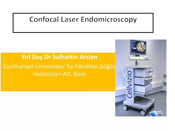
Confocal Laser Endomicroscopy. Yrd Doç Dr Sulhattin Arslan Cumhuriyet Üniversitesi Tıp Fakültesi Göğüs Hastalıkları AD, Sivas. HERHANGİ BİR KURUM YA DA ŞAHIS İLE BİR ÇIKAR İLİŞKİM YOKTUR. Confocal Laser Endomicroscopy.

E N D
ConfocalLaserEndomicroscopy YrdDoçDrSulhattin Arslan Cumhuriyet Üniversitesi Tıp Fakültesi Göğüs Hastalıkları AD, Sivas
HERHANGİ BİR KURUM YA DA ŞAHIS İLE BİR ÇIKAR İLİŞKİM YOKTUR
ConfocalLaserEndomicroscopy • Confocal endomicroscopes aim at providing microscopic imaging of a living tissue to the clinician • ‘‘Optical biopsies’’
ConfocalLaserEndomicroscopy • Such systems have recently been applied to the explorations of several organs, including the upper and the lower GIS, pancreato-biliary tract • More recently to the proximal and distal airways in vivo
ConfocalLaserEndomicroscopy 1- Optisan/Pentax Microendoscopy,Japan 2- CellVizio-LungR /Mauna Kea Tech, French Commercially;
Optisan/PentaxEndomicroscopy Optiscan endomicroscopy of the gastrointestinal tract provide imagesvery close to conventional histology
Optisan/PentaxEndomikroskopi Two potential drawbacks explainwhy this system is not yet available for the respiratory tractimaging; Largediameter of distal Scanning rate is tooslow (1 frame/second) Respiratorysystem is more mobile
CellVizio-LungR • A probe-based confocal laser endomicroscopy • Designed for imaging of the internal micro-structures of anatomical tissues
CellVizio-LungR The device has a laser scanning technology adapted to image by means of an optical fiber bundle
CellVizio-LungR Uses the principle of proximal scanning Carriesthelighttothescannedareaby fiber bundleandminiprobe Thelight delivery, scanning, spectral filtering, and imaging systemsare located at the proximal part of the device Thedistalpartincludes a connectorwhichtransferstheimagetotheminiprobeandthelaserscanningunit
ConfocalLaserEndomicroscopy • Confocalmicroscopyhelpstoimage in vivocellsandtissuesbyobtaininglateralandaxialresolution • Theilluminationanddetectionsystems being conjugated on the same focal plane, aretermedconfocal
ConfocalLaserEndomicroscopy • 488 nm wavelength is used for imaging • Microspectrometer experiments have clearly demonstrated thatthe main fluorescence signal emitted after 488 nm excitationfrom both bronchial and alveolar human systems originates fromthe elastin component of the tissue • Elastin fibers
ConfocalLaserEndomicroscopy • 1.4 nm diameter miniprobe • 488-nm-wavelength laser light • 9-12 frames per second • Depth of focus of 0 to 50 micron • Field of view of 600 3 600 micron • Lateral resolution of 3.5 μm • Up to 30,000 microfibers Histological quality image
MiniprobE • The miniprobe is applied onto thebronchial wall surface or advanced into a distal bronchiole down tothe acinusandobtainsimagesfromtheseanatomicalareas. Thebasementmembrane of the proximal airways Peripheralinterstitial Alveolarwall Macrophage Bronchialepitheliallayer Peripherallungnodules
ConfocalLaserEndomicroscopy Alveolar septal wall Microvessels Macrophage Basement membrane
ConfocalLaserEndomicroscopy • After the procedure Hemorrhage (because of miniprobe) there is no need to intervention
ConfocalLaserEndomicroscopy • Contrast agent Intravenous: Fluorescein %1,4 emesis, hypotension and epigastric pain Topical: Acriflavine, Metilen blue
Imaging of the Proximal Bronchi • Underlocalanesthesia • Miniprobe is introducedinto theworking channel of the bronchoscope • The depth of focus being 50 mm below the contactsurface, subepithelialconnectivetissue lamina densa, laminareticularis 480 nmfluoreskeine bronchialbasementmembrane Benign, malignant/premalignantbronchialalterations
FromtheDistalBronchioles Down to the Lung Acini Elastin represents up to 50% of the peripheral lung connective tissue fibers Bronchial wall, alveolus, extra-alveolar microvessels
Confocallaserendomicroscopyfordiagnosinglungcancer in vivo 32 patients with suspected malignancies underwent bronchoscopy with endomicroscopy using acriflavine 75,522 confocalimagesfrom 56 differentlocations Neoplasticchanges: Sensitivity, 96% Specificity, 87% Accuracy, 91% Fuchs et al. EurRespir J. 2012 Sep 20. [Epub ahead of print]
ConfocalLaserEndomicroscopy • Peripheral pulmonary nodule • Interstitial pathology • Bronchi and bronchial structure • Chronic rejection after lung transplantation
limitations • Can not obtaintissuesample • Onlyelastin fiber image