Download
1 / 50
530 likes | 902 Views
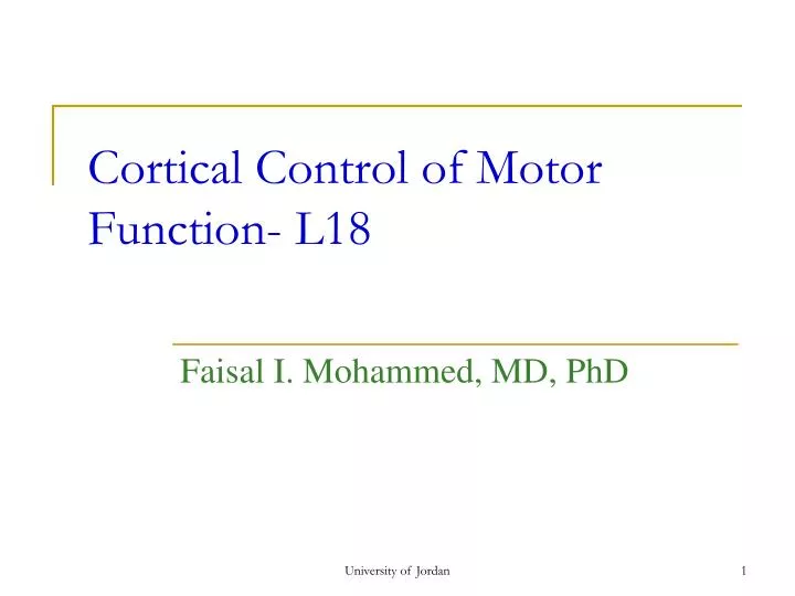
Cortical Control of Motor Function- L18. Faisal I. Mohammed, MD, PhD. Objectives. Recognize cerebral cortical motor areas Delineate the cortical control of the corticospinal pathways Interpret some of the cortical abnormalities. Red Nucleus. DSC & VSC. Red Nucleus. VA/VL Thalamus.

E N D
Cortical Control of Motor Function- L18 Faisal I. Mohammed, MD, PhD University of Jordan
Objectives • Recognize cerebral cortical motor areas • Delineate the cortical control of the corticospinal pathways • Interpret some of the cortical abnormalities University of Jordan
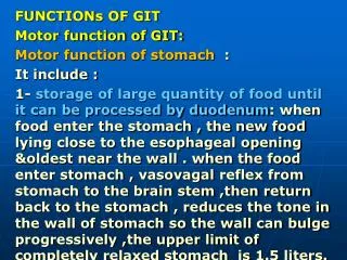
Red Nucleus DSC & VSC Red Nucleus VA/VL Thalamus Cerebral Cortex C.Spinal Rubrospinal B.G Spino-cerebellum Pontine Brain stem Centers Lateral Reticular Nucleus Inferior Olivary Nucleus Spinal Relay Nuclei Spinal Motor Centers Muscles Receptors Motor Command Feed Back Command Monitor Corrective Command Motor System
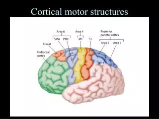
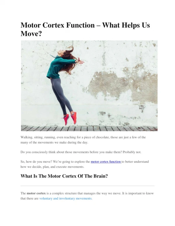
Motor Cortex • Divided into 3 sub areas • primary motor cortex • unequal topographic representation • fine motor movement elicited by stimulation • premotor area • topographical organization similar to primary motor cortex • stimulation results in movement of muscle groups to perform a specific task • works in concert with other motor areas University of Jordan
Motor Cortex (Cont.) • supplemental motor area • topographically organized • simulation often elicits bilateral movements. • functions in concert with premotor area to provide attitudinal, fixation or positional movement for the body • it provides the background for fine motor control of the arms and hands by premotor and primary motor cortex University of Jordan
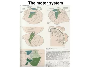
Motor Areas of the Cortex University of Jordan
Functional organization of the primary Motor Cortex • Located in the precentral gyrus of the frontal lobe. • More cortical area is devoted to those muscles involved in skilled, complex or delicate movements, that have more motor units i.e the cortical representation is proportional to the No of motor units University of Jordan
Specialized Areas of the Motor Cortex • Broca’s area • damage causes decreased speech capability • closely associated area controls appropriate respiratory function for speech • eye fixation and head rotation area • for coordinated head and eye movements • hand skills area • damage causes motor apraxiathe inability to perform fine hand movements University of Jordan
Transmission of Cortical Motor Signals • Direct pathway • corticospinal tract • for discrete detailed movement • Indirect pathway • signals to basal ganglia, cerebellum, and brainstem nuclei University of Jordan
Corticospinal Fibers • 34,000 Betz cell fibers, make up only about 3% of the total number of fibers • 97% of the 1 million fibers are small diameter fibers • conduct background tonic signals • feedback signals from the cortex to control intensity of the various sensory signals to the brain University of Jordan
Corticospinal pathways University of Jordan
Other Pathways from the Motor Cortex • Betz collaterals back to cortex sharpen the boundaries of the excitatory signal • Fibers to caudate nucleus and putamen of the basal ganglia • Fibers to the red nucleus, which then sends axons to the cord in the rubrospinal tract • Reticular substance, vestibular nuclei and pons then to the cerebellum • Therefore the basal ganglia, brain stem and cerebellum receive a large number of signals from the cortex. University of Jordan
Incoming Sensory Pathways to Motor Cortex • Subcortical fibers from adjacent areas of the cortex especially from somatic sensory areas of parietal cortex and visual and auditory cortex. • Subcortical fibers from opposite hemisphere which pass through corpus callosum. • Somatic sensory fibers from ventrobasal complex of the thalamus (i.e., cutaneous and proprioceptive fibers). University of Jordan
Incoming Sensory Pathways to Motor Cortex (Cont.) • Ventrolateral and ventroanterior nuclei of thalamus for coordination of function between motor cortex, basal ganglia, and cerebellum. • Fibers from the intralaminar nuclei of thalamus (control level of excitability of the motor cortex), some of these may be pain fibers. University of Jordan
Sensory Feedback is Important for Motor Control • Feedback from muscle spindle, tactile receptors, and proprioceptors fine tunes muscle movement. • Length mismatch in spindle causes auto correction. • Compression of skin provides sensory feedback to motor cortex on degree of effectiveness of intended action. University of Jordan
Excitation of Spinal Motor Neurons • Motor neurons in cortex reside in layer V. • Excitation of 50-100 giant pyramidal cells is needed to cause muscle contraction. • Most corticospinal fibers synapse with interneurons. • Some corticospinal and rubrospinal neurons synapse directly with alpha motor neurons in the spinal cord especially in the cervical enlargement. • These motor neurons innervate muscles of the fingers and hand. University of Jordan
Final Common Pathway University of Jordan
Lesions of the Motor Cortex • Primary motor cortex - loss of voluntary control of discrete movement of the distal segments of the limbs. • Basal ganglia - muscle spasticity from loss of inhibitory input from accessory areas of the cortex that inhibit excitatory brainstem motor nuclei. University of Jordan
Thank You University of Jordan
Cerebral Cortex; Intellectual Functions of the Brain; Learning and Memory- L22- L23 Faisal I. Mohammed, MD, PhD University of Jordan
Objectives • Describe intellectual functions of the cerebral cortical areas • Explain memory and learning • Outline the dominant and non-dominant hemispheres • Delineate language areas of speech University of Jordan
Physiologic Anatomy of Cerebral Cortex • Each area of the cortex is connected to a specific part of the thalamus. • When thalamic connection is lost cortical function stops. • All sensory pathways pass through the thalamus with the exception of some olfactory signals.
Physiological Anatomy of the Cerebral Cortex University of Jordan
Dominant and Non-Dominant Hemisphere • Wernicke’s area more developed in one hemisphere, responsible for verbal symbolism and related intelligence. Dominance is related to Language • 95% of population has a left dominant hemisphere. • Wernicke’s area can be as much as 50% larger in the dominant hemisphere.
Dominant and Non-Dominant Hemisphere (Cont’d) • Damage to dominant Wernicke’s area leads to dementia. • Non-dominant side related to other forms of sensory intelligence (music, sensory feelings).
Hemispheric Lateralization University of Jordan
Language Areas • Located in a large area surrounding the left (or language-dominant) lateral sulcus • Major parts and functions: • Wernicke’s area – involved in sounding out unfamiliar words –sensory aspect of speech- damage sensory aphasia (Receptive aphasia) • Broca’s area – speech preparation and production- motor aspect of speech – damage motor aphasia (expressive aphasia0 • Both Wernicke’s and Broca’s area damage global aphasia • Lateral prefrontal cortex – language comprehension and word analysis • Lateral and ventral temporal lobe – coordinate auditory and visual aspects of language University of Jordan
Brain Organization and Handedness • Close to 90% of people are right-handed and close to 10% are left-handed and a small number are ambidextrous (use both hands) • 95% of right-handers process speech primarily in the left hemisphere • left-handers: around 65% in left hemisphere, 15-20% in right hemisphere, 15-20% in both • More than 90% of people are born with the left hemisphere area that controls the movement of the right hand is bigger • They tend to use the right hand, this area grows and become dominant University of Jordan
Brain Organization and Handedness …cont • Left handed people have their right cerebral hemisphere area that controls the movement of the left hand bigger • If they use the left hand then this area grow and become dominant • They still can convert and the younger the more easier if they start to use their right hand instead and then they become right handed • Same applies for using the legs University of Jordan
Intellectual Functions of the Prefrontal Association Area • responsible for calling forth stored information and using it to obtain a goal • responsible for concerted thinking in a logical sequence • damage causes an inability to keep tract of simultaneous bits of information, easily distracted
Intellectual Functions of the Prefrontal Association Area (Cont’d) • elaboration of thought • prognosticate, plan, consider consequences of motor actions before they are performed • correlate widely divergent information, control one’s activities • Personality trait and behavior that confines to values and manners of the culture
Pathways for Auditory Communication 5. activation of motor programs in Broca’s area for control of word formation 6. transmission of signals to motor cortex to control speech muscles 4. transmission via the arcuate fasciculus to Broca’s area 3. formation of the word that expresses a particular thought 2. interpretation of the word and the thought that the word expresses in Wernicke’s area 1. primary auditory area recognition of the sound as a word University of Jordan
Pathways for Visual Communication 5.transmission of signals to motor cortex to control speech muscles 4. then to Broca’s area for motor formation of the word 3. visual input reaches full level of interpretation in Wernicke’s area 2. processing of the visual information in the parietal-temporal-occipital association cortex, the angular gyrus region 1. receive the visual input in primary visual area
Wernicke's aphasia Sensory Aspects of Communication Destruction of the visual and auditory association areas results in an inability to understand the written or spoken word. University of Jordan
Motor Aspects of Communication • Speech involves two things • formation in the mind of thoughts to be expressed and the choice of words • motor control of vocalization and the act of vocalization • Formation of word, thought and choice of words is function of Wernicke’s area. • Broca’s area controls the motor coordination required for speech.
Function of the Corpus Callosum • connects the two hemispheres and allows transfer of information • interruption of these fibers can lead to bizarre types of anomalies • dominant hemisphere understands spoken word • non dominant hemisphere understands written word and can elicit motor response without dominant side knowing why response was performed
Thoughts and Memory • Neural mechanism for thought is not known. • Most likely a specific pattern of simultaneous neural activity in many brain areas. • Destruction of cerebral cortex does not prevent one from thinking. • However, depth of thought and level of awareness may be less.
Memory and learning • Learning is acquiring new sensory information or motor skills • Change in the capability of synaptic transmission from neuron to neuron as a result of prior stimulation. • Memory trace is a specific pattern or pathway of signal transmission. • Once established they can be activated by the thinking mind to reproduce the pattern and thus the memory.
3 Types of Memory • immediate memory • lasts for seconds or minutes (remembering 10 digit phone number) • short-term memory • lasts for days to weeks • long-term memory • lasts for years or for a lifetime
Mechanism of Memory • Immediate memory may result from synaptic potentiation through the accumulation of calcium in the presynaptic membrane. • would promote neurotransmitter release • Short-term memory may result from a temporary physical or chemical change in the pre- or postsynaptic membrane.
Cellular Basis for Memory • repetitive stimulation causes a progressive decline in sensitivity called habituation • results from progressive decline in the number of active calcium channels • less calcium entry less transmitter released • stimulation of facilitator terminal prevents habituation
Molecular Basis for Memory Transmitter activates G protein which in turn activates adenylate cyclase resulting in an increase in cAMP. cAMP activates a protein kinase that phosphorylates a component of the K+ channel blocking its activity. This prolongs the action potential which increases transmitter release. Figure 57-9; Guyton & Hall University of Jordan
Long-Term Memory • results from a structural change in the synapse • increase in the area for vesicular release therefore, more transmitter is released • during periods of inactivity the area decreases in size • enlargement of the release site area results from synthesis of release site proteins
Consolidation of Memory • converting immediate into short and long-term memory • results from chemical, physical and anatomical changes in the synapse • requires time • interruption of the process by electrical shock or by anesthesia will prevent memory development • rehearsal enhances consolidation
Brain Centers and Memory • Thalamic structures are important for recalling memories. • Damage to thalamus causes retrograde amnesia or the inability to recall stored experiences. • Thalamus scans the cortex for the area and the circuit for the stored memory.
Thank You University of Jordan