Download
1 / 21
260 likes | 812 Views
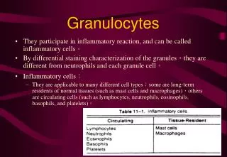
Granulocytes. Our bodies make 80 million granulocytes every minute! They are short-lived and survive for only 2-3 days. 60-70% of all leucocytes in blood are granulocytes. They are also called polymorphonuclear cells (PMN ). They can be distinguished by their appearance under Write stain into:.

E N D
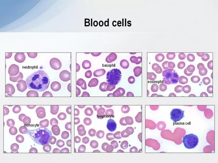
Granulocytes Our bodies make 80 million granulocytes every minute! They are short-lived and survive for only 2-3 days. 60-70% of all leucocytes in blood are granulocytes. They are also called polymorphonuclear cells (PMN). They can be distinguished by their appearanceunder Write stain into:
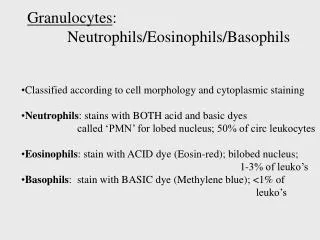
Neutrophils • ~90% of all granulocytes • Multi-lobed nucleus • 10-20μm in diameter • Phagocytose microbes and digest them • Degranulate following activation via FcR • Primary (azurophilic) granules: • acid hydrolases, myeloperoxidase, • lysozyme • Secondary (specific) granules: • lactoferrin, lysozyme • Respond to chemotactic signals such as complement fragments (C5a), kinins, fibrinolytic products
Neutrophils - Primary granules Primary granules of neutrophils contain cationic proteins and defensins that are used to kill bacteria, proteolytic enzymes and cathepsin G to breakdown bacterial proteins, lysozyme to break down bacterial cell walls, and myeloperoxidase that used to generate toxic bacteria-killing substances. In addition, secretions from the primary granules of neutrophils stimulate the phagocytosis of IgG antibody-coated bacteria. The secondary granules contain compounds that are involved in the formation of toxic oxygen compounds, lysozyme, and lactoferrin that used to take essential iron from bacteria.
Basophils - Mast cells • 0.2% of leucocytes • Deep violet blue granules • Granules contain histamine, heparin, SRS-A and ECF-A, peroxidase (basophils), acid and alkaline phosphatases (mast cells) • Degranulation following antigen binding to IgE bound on cell surface by FcεR
Eosinophils • 2-5% of leucocytes • Granules are membrane-bound organelles, fuse. with plasma membrane upon binding to parasite via FcR (γ or ε). • professional antigen presenting cells. • Granules contain peroxidase, acid phosphatase, and cationic proteins (e.g cathepsin). • Involved in the destruction of tumor cells, and promote the repair of damaged tissues. • Respond to chemotactic signals from T cells (ECF), mast cells and basophils.
Anti-infectious response of PMNs • Chemotaxis and migration to the extra-vascular compartment: • Interact with endothelial cells via selectins. • Chemotactic factors are of bacterial origin (e.g. fMLF), tissue necrosis (leukotriene B4), monocyte/lymphocye activation (e.g. IL-8, RANTES), by-products of complement (C5a). • Cell activation, express CAMs (integrin of CD11/CD18). • Bind to platelet endothelial cell adhesion molecule 1 (PECAM-1), which mediates the process of diapedesis.
Anti-infectious response of PMNs 2. Phagocytosis • The recognition of infectious agents (mainly opsonized particles), via two types of receptors: • FcγR: allows the recognition of antibody-coated particles. • CR1: expressed on all phagocytic cells.
3. Intracellular killing Anti-infectious response of PMNs a. Oxygen - dependent killing “Respiratory burst” - triggered by binding of microorganism to macrophage Glucose + NADP+ hexose monophosphate pentose phosphate shunt + NADPH NADPH + O2 cytochrome b-245 NADP+ + O2- (superoxide) 2O2- + 2H+ H2O2 + O2 spontaneous dismutation O2- + H2O2.OH + OH- + O2 H2O2 + Cl- myeloperoxidase OCl- + H2O OCl- + H2O O2 + Cl- + H2O
Anti-infectious response of PMNs b. Oxygen - independent killing
Mononuclear phagocytes “Fixed” macrophages Spleen - Macrophages attached to sinusoids, free within sinusoids and in red pulp. Lymph nodes - Macrophages line the sinuses, are found in the lumen of the sinuses, in the trabeculae and within the germinal centres. Liver - Kupffer cells line the hepatic sinusoids. Bone marrow - bone marrow macrophages are most numerous adjacent to venous sinusoids and amongst groups of erythroblasts. Kidney - Mesangial cells populate the mesangia of the glomeruli Brain - Microglial cells are the macrophages of the CNS and are mesodermal and not ependymal in origin Bone marrow - Osteoclasts are large multinucleated giant cell macrophages in the bone marrow. Skin - Langerhans cells of the epidermis “Wandering” macrophages Lung- Alveolar macrophages Gut - Serosal macrophages Body cavities - e.g. peritoneal macrophages Blood - Monocyte Connective tissue - Histiocytes
Receptors on Macrophages Mannosyl-fucosyl receptors - bind to sugars on the surface of microorganisms and effete cells such as lymphocytes CD14 - lipopolysaccharide receptor Toll receptors - receptors for lipopolysaccharide and microbial DNA Fc-receptors - bind to immunoglobulin bound to the surface of the microorganism triggering phagocytosis and cell killing Complement receptors - bind to complement fixed to the surface of the microbe MHC class II - antigen presentation to T cells
Roles of macrophages Cytokines secreted by macrophages TNFα IL1-b IL-6 IL-10 IL-12 Cytokines acting on macrophages IFNγ IL-4 IL-13 TGF- β M-CSF Chemokines acting on macrophages RANTES MCP-1 MCP-3
Roles of macrophages Microbicidal activity oxygen dependent H2O2, .O2-, NO Modulation of the immune response IL-12 - Th1 oxygen independent lysozyme, acid hydrolases cationic proteins IL-10 - Th2 Lymphocyte activation Inflammation and fever antigen processing antigen presentation IL-1 production IL-6, TNF-α, IL-1 prostaglandins complement clotting factors Tissue reorganisation elastase, collagenase Hyaluronidase fibroblast stimulating factors angiogenesis factors