Download
1 / 82

E N D
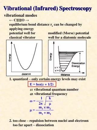
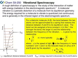
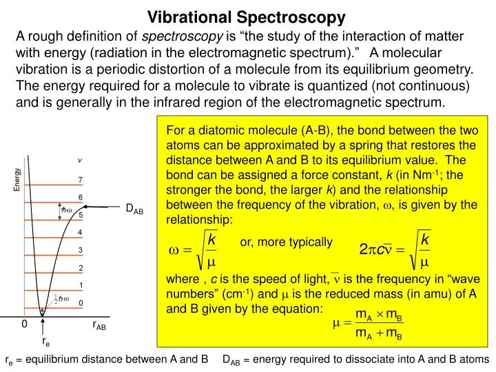
Vibrational Spectroscopy A rough definition of spectroscopy is “the study of the interaction of matter with energy (radiation in the electromagnetic spectrum).” A molecular vibration is a periodic distortion of a molecule from its equilibrium geometry. The energy required for a molecule to vibrate is quantized (not continuous) and is generally in the infrared region of the electromagnetic spectrum. For a diatomic molecule (A-B), the bond between the two atoms can be approximated by a spring that restores the distance between A and B to its equilibrium value. The bond can be assigned a force constant, k (in Nm-1; the stronger the bond, the larger k) and the relationship between the frequency of the vibration, , is given by the relationship: DAB or, more typically where , c is the speed of light, is the frequency in “wave numbers” (cm-1) and is the reduced mass (in amu) of A and B given by the equation: 0 rAB re re = equilibrium distance between A and B DAB = energy required to dissociate into A and B atoms
Infrared Radiation Portion of the electromagnetic spectrum between visible light and microwaves full range for IR is 10000-400 cm-1 of importance here is 4000-400 cm-1 (wavenumbers) or 2.2-25 mm (wavelength) Note: cm-1 is proportional to Energy cm-1 = 104/mm this energy is absorbed by molecules and converted to molecular vibration
IR Absorption IR absorptions are characteristic of entire molecule or essentially a molecular fingerprint vibration spectrum appear as bands molecular vibration is not a single energy as also depends on molecular rotation band intensities expressed as either transmission (T) or absorption (A) A = log10(1/T)
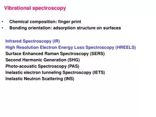
Molecular Vibrations Stretching is a rhythmical movement along a bond Bending is a vibration that may consist of a change in bond angle (twisting, rocking and torsional vib.) Vibrations that result in change of dipole moment give rise to IR absorptions alternating electric field produced by changing dipole couples the molecular vibration to the oscillating electric field of the radiation
Vibrations for H2O and CO2 3650 cm-1 3756 cm-1 1596 cm-1 Symmetrical asymmetrical scissoring stretch stretch (inactive in IR) for CO2 1340 cm-1 2350 cm-1 666 cm-1 + - +
Bending for CH2 + + - Asymmetric symmetric in-plane out-of-plane stretch stretch bend bend 2926 cm-1 2853 cm-1 1465 cm-1 1350-1150 cm-1 - + In plane bend or rocking 1350-1150cm-1 Out-of plane bend or twist 1350-1150cm-1
Assignments of Bands For a stretching frequency interruption based on Hooke’s Law: Frequency = 1/2pc[(k/(MxMy/Mx+My)]1/2 where f = force constant of bond and M is mass f is about 5 x10⁵ dyne/cmfor single bond 2x that for double bond and 3x that for triple bond C-H stretch:calc: 3040 cm-1actual CH3: 2960-2850 cm-1 Note: for C-D: stretch is 21/2 x that of C-H
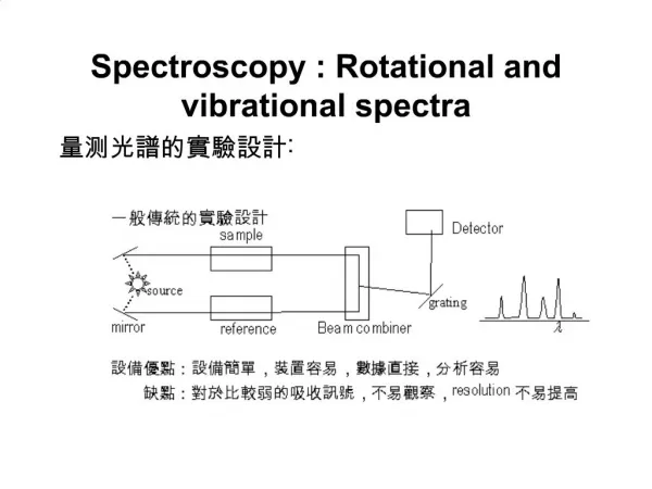
Instrumentation Requirements: source of IR radiation, sample, detector
Sample Handling IR spectra can be obtained for gases, liquids and solids Liquids: may be neat or in solution Neat: between to NaCl plates (0.01 mm film) (NaCl does not absorb until 600 cm-1) thick samples absorb too strongly: poor spectrum Solution: cells are 0.1-1 mm thick(0.1-1 mL in volume) requires second cell of pure solvent to correct for absorptions of solvent Solids: usually as a mull (supension) in nujol oil (free of IR absorptions 4000-250 cm-1 or dispersed in KCl pellet
Spectral Interpretation Precise and complete interpretation is NOT possible thus must use IR in conjunction with other techniques but Functional group region: 4000-1300 cm-1: eg: OH, NH, C=O, S-H, CºC Many functional groups exhibit characteristic bands Fingerprint regions: 1300-650 cm-1absorptions here are usually complex some interpretation is possible similar compounds give similar spectra but fingerprint is unique
An Organic Example CºN stretch 2226 Aromatic C-H bands
Nuclear Magnetic Resonance Sample in a magnetic field absorbs radio frequency radiation absorption depends on certain nuclei in molecule initially we deal with 1H (proton) NMR inspection of NMR provides much more structural data than MS or IR
Magnetic Nuclei Nuclei with odd mass, odd atomic number or both have quantized spin angular momentum eg 11H, 21H, 136C, 147N, 3115P spin quantum number, I = 0, 1/2, 1, 3/2 ….. For 11H,136C,3115P: I = 1/2 For 21H, 147N I = 1 (nonspherical charge distribution: electric quadrupole) number of states in magnetic field 2I+1
In a Magnetic Field DE=(hn/2p)Bo Bo is related to strength of magnetic field h is Planck’s Constant DEis in the radio frequency range
Absorbance of RF In magnetic field spinning nucleus precesses about applied magnetic field (Larmor Frequency) when same frequency RF is applied electric field of radiation and electric field of precessing nucleus couple E is transferred and spin changes -Resonance
Relaxation How is this energy dissipated? T1 spin-lattice or longitudinal relaxation process transfer of E from excited protons to surrounding protons T2 spin-spin transverse relaxation transfer of E among precessing protons, result is line broadening
Instrumentation Magnetic field, radio frequency generator
Instrument 1945-46 at Stanford Professor Bloch Nobel Prize 1952
Sample Typically if want to observe 1H NMR need to avoid solvent with protons used deuterated solvent or solvent with no protons for example: C6D6, CDCl3 or CCl4 sample is held in a 5mm tube typically 2 mg in 0.5 mL) sample is spun in the magnetic field to average out field inhomogeneities
Magnets 1953: 1.41 Tessla or 60 MHz for proton resonance Now: 200-500 MHz magnets are common as high as 900 MHz in some NMR research Labs magnetic fields are large: in the case of 500 MHz magnetic 5Gauss lines forma a 15 ft sphere about the magnets
Chemical Shift Electron density in a magnetic field circulates generating a magnetic field in opposition to the applied field thus shielding the nucleus…. Since electron density for each type of proton environment is different get different resonance absorption of RF neff = (g/2p)Bo(1-s) s is the shielding constant reference position relative to the standard TMS tetramethylsilane
NMR Scale Set TMS to zero Hz (300 MHz magnet) if we use this scale must specify the strength of magnet as frequency of resonance will change with field better to use dimensionless units: d(ppm) freq/applied field x 106 = d 0 Hz 3000 Hz 0 ppm 10 ppm
NMR Scales 3000 Hz 300 MHz 0 Hz 0 ppm 10 ppm 6000 Hz 600 MHz 0 Hz 10 ppm 0 ppm
Field Strength Effect 60 MHz 300 MHz
Chemical Shifts As the shift depends somewhat on electron density electronegativity may be a guide for chemical shifts electron density around protons of TMS is high positive d increases to left of TMS increase d means deshielded relative to TMS since C is more electronegative than C expect: R3CH>R2CH2>RCH3>CH4 1.6 1.2 0.8
NMR Scales 3000 Hz 300 MHz 0 Hz 0 ppm 10 ppm Higher frequency-less shielded Lower frequency-more shielded 6000 Hz 600 MHz 0 Hz 10 ppm 0 ppm
Acetylene based on electronegativity expect higher chemical shift than ethylene Apparent anomaly H-CºC chemical shift is 1.8 ppm WHY? linear molecule: if aligned with magnetic field then p-electrons can circulate at right angles to field and generate magnetic field in opposition to applied field thus: protons experience diminished field and thus resonance at lower frequency than expected 1.7-1.8 ppm
Aldehydes Deshielded position of aldehyde proton observed at 9.97 ppm (acetaldehyde)
Benzene Ring current effect deshields aromatic protons 7.0-8.0 ppm (depending on substitution)
[18]Annulene Outside protons are deshielded 9.3 ppm protons on inside shielded -3.0 ppm
Acetophenone All protons are deshielded due tp ring currents Ortho-protons are further deshielded due to carbonyl meta, para 7.40 ppm ortho 7.85 ppm Ring current effect infer planarity and aromaticity
General Regions of Chemical Shifts alkyne monosubstituted aliphatic disubstituted aliphatic alkene Aromatic aldehydic ppm 10 9 8 7 6 5 4 3 2 1 0
Integration: Benzyl Acetate Integration 5:2:3 At high resolution see multiplet
Spin-spin Coupling Chemically inequivalent protons: field of one proton affects the other normally only see up to 3-bond coupling -1/2 -1/2 +1/2 +1/2
Spin-spin Coupling Each proton has a unique absorption but effected by magnetic field of other proton J is the coupling constant
Coupling C-H sees CH2 protons CH2 sees C-H proton (+1, 0, -1) (+1/2, -1/2)
Ethylbenzene Typical ethyl pattern A2B3 triplet quartet
Ethanol in CDCl3 Rapid exchange of OH: do not see coupling CH3CH2OH
Ethanol in DMSO No exchange CH3CH2OH
Doublet of Quartets CH3CH2OH Can see: J(CH2-OH) and J(CH3-CH2)
N-methylcarbamate 14N has I =1, if exchange is rapid no coupling intermediate or slow --broad NH;
H-C-N-H Coupling In trifluoroacetic acid, amine is protonated see methylene coupling to N-H protons
Fluoroacetone, CH3COCH2F 19F has I = 1/2 J2 J4
Other Magnetic Heteroatoms 2H (Deuterium): I = 1; simplifies proton spectrum as H-D coupling is small X-CH2-CH2-CH2-COY X-CH2-CH2-CD2-COY triplet, quintet, triplet triplet, slightly broad triplet 31P: I = 1/2 (100% natural abundance) large coupling constants P-H 200-700 Hz 29Si: I = 1/2 (4.7% Natural abundance) Si-CH 6 Hz; low intensity (satellites) 13C: I = 1/2 (1.1% Natural abundance) not seen unless enriched with 13C
Chemical Shift Equivalence Nuclei are chemical shift equivalent if they are interchangeable through a symmetry operation or by a rapid process. Rotation about a simple axis (Cn) Reflection through a plane of symmetry (s) Inversion through a center of symmetry (i)