Download
1 / 19
270 likes | 544 Views
Calmodulin Reaches Out. Yubin Zhou. Calmodulin: the intracellular calcium sensor . A versatile and ubiquitously expressed EF-hand Ca 2+ -binding protein in eukaryotes (~8.8 µM). . The first structure determined in 1980’s; Highest resolution with 1 Å.

E N D
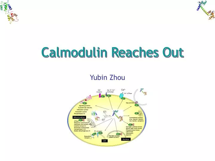
Calmodulin Reaches Out Yubin Zhou
Calmodulin: the intracellular calcium sensor • A versatile and ubiquitously expressed EF-hand Ca2+-binding protein in eukaryotes (~8.8 µM). • The first structure determined in 1980’s; Highest resolution with 1 Å. • First discovered in the brain and heart in 1960’s. • Regulates the activity of a large array of different target molecules (>300). • Small (148 a.a., ~17 KDa) and acidic (pI: ~4.0). • Two autonomous lobes: “low-affinity” (~2 µM) N-terminal domain and “high-affinity” (~0.2 µM)C-terminal domain • Mg2+ and K+ also bind to CaM, but with 1000-10000 folds lower affinity Black D. et al. Cell calcium 2004 (35): 415-425
7 1 5 3 12 9 Calmodulin: a prototypical EF-hand protein E n * * n n * * n E n * * n n * * n n * * n n * * n * * n n * * 1 x Ca 1 x * =O Ca * * 4 Y 3 Y z 14 G * bi z 12 * 6 Z x 11 * =O n * y9 5 Z bi * * * G x9 y7 n * =O H20 =O =O Canonical EF-hand Ca2+-binding motif Pseudo EF-hand Ca2+-binding motif
Ca2+ Na+/K+-Ca2+ exchanger EGF receptor CaM in action Ca2+ ATPase CaM CaM CaM G E Gap junction CaM CaM Signaling proteins G-protein βγ subunits Adenylate cyclase Ca2+ Guanylate cyclase Phospholipase A2 CaM Mitochondrion Cytoskeletal proteins (e.g., Myosin, tubulin, tau, dynein, synapsin) Ca2+ E CaM ATPase Ubiquinol cytochrome c reductase core protein 2(?) Creatine kinase (?) Glutamate oxaloacetate transaminase 2 (?) Malate dehydrogenase (?) E CaM Glycogen synthase kinase Myosin light chain kinase Phosphorylase kinase CaM kinase Calcineurin Spectrin IP3 receptor CaM E CaM CaM P68 RNA helicase Ryanodine receptor hnRNP A2 and C Nucleus ER Yang, J. Calcium Binding Proteins, 2006
Ca What confers its recognition diversity? Conformtional plasticity Ca2+-free Calmodulin (1cfc) Ca2+-binding Calmodulin (3cln) Calmodulin:peptide complex (1cdl) Varied Ca2+-binding affinities between N and C lobes Exposable hydrophobic cleft b/w helices Flexible tethering linker region (78-81): tumbling motion
CaM: apo form and Ca2+-loaded form Apo-CaM: closed state two sets of four-helix bundle Ca2+-CaM: open state Dumbbell shape Exposure of hydrophobic clefts Interhelical angles in CaM I-II III-IV V-VI VII-VIII Apo (NMR) 50 48 38 53 Ca2+ (NMR) 76 78 70 78 Ca2+ (X-ray) 92 93 75 85 Chou, J et al. Nature SB, 2001 (8): 990-997
Ca2+-induced exposure of hydrophobic core Ikura M, et al. PNAS 2006 (103): 1159-1164
Ca2+-induced conformational changes Shen, Y et al. EMBO, 2005; 24: 929-941
Cooperative Ca2+-binding process nHill=1.8~2.5 unpublished data
How to obtain site-specific Ca2+-binding constants • Mutational studies more than 50 mutations made on the loop, helices, linker region and hydrophobic region. • Peptide models Easy synthesis; less defined structure. • Proteolytic fragmentation Eliminate the long distance cooperativity that occurs b/w two lobes. • Grafting approach Eliminate the contribution of conformational change; Obtain intrinsic Ca2+-binding affinities; Estimate cooperativity. Yang, J et al. Protein and Peptide Letters. 2003 (10): 331-345
Diversity in Activation Mechanisms Autoinhibitory domain (AID) displacement CaM wraps around the helical AID, enclosing it in a hydrophobic channel within the globular core (e.g., CaM kinase II, MLCKs, CaMKK, calcineurin) N C Active site remodeling CaM induces the rearrangement of the target and creates an active site that is otherwise totally exposed to the solvent (e.g., anthrax adenylyl cyclase). CaM only binds two Ca2+ at its C-terminal high-affinity lobe. (e.g., anthrax adenylyl cyclase or edema factor) N C CaM-induced oligomerization CaM is constitutively associated with the and forms a 2:2 complex. C-terminal lobe tethers the channel whereas the N-terminal lobe (binds Ca2+) induces the dimerization, thus coupling the changes in [Ca2+]i with membrane potential. (e.g., small conductance Ca2+-activated K+ channel (SK) C C C N N N N C Hoeflich, K et al. CELL, 2002; 108: 739-742
A typical CaM/peptide complex • Collapsed compact structure • Central helix: unwound and bend by 100o and to rotate by 120o • C- and N-terminal lobes are brought close together • The interaction is largely driven by the hydrophobic interaction involving Met in CaM • C-terminal end has a negatively charged rim • N-terminal patch has clusters of negatively and positively charged residues CaM/CaMKIIalpha complex
Prediction of CaM recognition sequences Common structural features: • Basic, amphiphilic alpha-helix; • Large hydrophobic residues in conserved positions: 1-8-14; 1-5-10 etc; Criteria to judge a potential CaM recognition sequence: Average hydrophobicity Average hydrophobic moment Average propesity for alpha-helix formation Percent of hydrophobic residue Net charge Number of basic residues Number of acidic residues http://calcium.uhnres.utoronto.ca/
Prediction of CaM recognation sequences Rhoads, A et al. FASEB, 1997; 11: 331-340
Prediction of CaM recognition sequences Yap K et al. JSFG, 2000; 1: 8-14
Prediction of CaM recognition sequences (example) Extracellular 41 72 178 208 Plasma membrane TM1 TM2 TM3 TM4 158 236 Intracellular 94 24 257 292 365 NH2 COOH 368 262 372 C Score 13699999999999999999631 Cx43 136 KYGIEEHGKVKMRGGLLRTYIIS 158 Cx44 127 APVRDDRGKVRIAGALLRTYVFN 149 Cx50 139 SSSSKGTKKFRLEGTLLRTYVCH 161 *::: * *****:: xxxxxxxxB#Bxxxxx#Bxxxxx 1 8
Conclusions and perspectives • A versatile and ubiquitously expressed EF-hand Ca2+-binding protein in eukaryotes. • It binds 4 calcium ions with differed affinites in a cooperative fashion • Its unique structural feature enables its interaction with more than 300 target molecules • At least three modes of interaction have been discovered: AID displacement, active site remodeling, induction of oligomerization • Biocomputational prediction of CaM recognition sequence is possible • Novel CaM-target interacting modes to be anticipated