Download
1 / 43
430 likes | 540 Views
Cell Cycle & Mitosis. Ms. Levensailor. Plan for understanding the cell cycle. Establish important vocabulary. Develop an understanding of the overall process. Dive into the details of the mitotic cell cycle. Know what is happening at each step.

E N D
Cell Cycle & Mitosis Ms. Levensailor
Plan for understanding the cell cycle • Establish important vocabulary. • Develop an understanding of the overall process. • Dive into the details of the mitotic cell cycle. • Know what is happening at each step. • Identify significant features of the cell during each step.
Cell Division Functions • Reproduction: asexual and sexual • Growth & Development: fertilized egg • Tissue renewal: repair and replacing cells that die • Involves the distribution of identical genetic material (DNA) to 2 daughter cells.
Cell Division • Genome: a cells DNA (genetic information). • Eukaryotic genomes are enormous. • DNA is packaged into chromosomes. • This packaging makes replication manageable within the cell. • Eukaryotes have a set number of chromosomes in each cell nucleus. • Human somatic cells (body cells not including reproductive cells) contain 46 chromosomes.
Cell Division • Each chromosome is one very long, linear DNA molecule. • Represents thousands of genes (an organisms inherited traits). • DNA is associated with proteins that maintain the structure of the chromosomes. • Chromatin: DNA-protein complex. • Organized into a long thin fiber.
Chromatin • After DNA is duplicated for division, chromatin is condensed! • It becomes densely folded and coiled. • We can now see it using a light microscope.
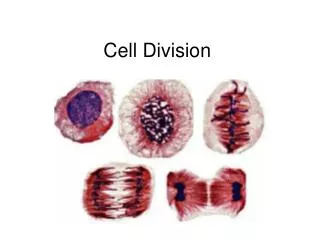
Sister Chromatids • Each duplicated chromosome has 2 sister chromatids. • Contain identical copies of the chromosome’s DNA molecule. • Centromere: narrow waist of the chromosome. • Pulled apart and repackaged as complete chromosome sets. • In 2 new nuclei, one at each end of the cell.
Mitosis • Mitosis: division of the nucleus. • Followed by cytokinesis. • Cytokinesis: division of the cytoplasm.
Details of the Cell Cycle • 2 main phases: • Interphase: • Accounts for 90% of the cycle. • Cell grows and copies chromosomes in prep for division. • Mitotic phase: • Includes mitosis and cytokinesis. • Shortest part of cell cycle.
Interphase • Divided into subphases: • G1 phase (first gap): • S phase (synthesis of DNA): Chromosomes are duplicated! • G2 phase (second gap): • During all 3 phases cell is growing by producing proteins and cytoplasmic organelles. *NOTE: Fill in graphic organizer!
Interphase • Nucleus is well defined and bound by the nuclear envelope. • 2 centrosomes are outside the nucleus. • Features a pair of centrioles (animal cells only). • Asters: microtubules extend from the centrosomes in radial arrays.
Interphase Onion roottip
Mitosis • Broken up into 5 main subphases: • Prophase • Prometaphase • Metaphase • Anaphase • Telophase
Prophase • Changes in the nucleus: • Chromatin fibers become more tightly coiled. • Condense into discrete chromosomes. • Chromosomes appear as 2 sister chromatids. • Nucleoli disappear. • Changes in the cytoplasm: • Mitotic spindle forms (made of microtubules). • Centrosomes move away from each other.
Prometaphase • Nuclear envelope fragments. • Microtubules interact with the chromosomes: • Bundles of microtubules extend from each pole and toward the middle of the cell. • Each of the chromatids has a kinetochore. • Located at the centromere. • Microtubules attach at the kinetochore.
Prophase Onion roottip
Metaphase • Centrosomes are at opposite poles of the cell. • Chromosomes line up on the metaphase plate. • Imaginary plane that is equidistant between the 2 poles. • Kinetochores are attached to microtubules of the opposite pole. • Spindle is formed. • Entire apparatus of microtubules.
Anaphase • Paired centromeres of each chromosome separate. • Frees sister chromatids from each other. • Now considered chromosomes. • Chromosomes move toward opposite poles. • Result of kinetochore microtubules.
Telophase • Daughter nuclei form at the two poles of the cell. • Nuclear envelope forms (from parent cell). • Chromatin fiber of each chromosome becomes less tightly coiled. • Mitosis is complete! • The division of one nucleus into 2 genetically identical nuclei.
Cytokinesis • Division of the cytoplasm. • 2 daughter cells appear at the end of mitosis. • In animal cells cytokinesis involves: • Formation of a cleavage furrow. • Pinching the cell in 2. • In plant cells: • No cleavage furrow. • Produce a cell plate.
Cytokinesis in Plants • During telophase: • Vesicles from the golgi apparatus move along microtubules to the middle of the cell. • This produces a cell plate. • Cell wall materials carried in the vesicles collect in the cell plate. • Cell plate enlarges and fuses with the plasma membrane. • Results in 2 daughter cells.
Mitotic Spindle • Mitosis is dependent on the mitotic spindle! • Consists of fibers made of microtubules and associated proteins. • Interesting note: while mitotic spindle is forming the microtubules of the cytoskeleton disassemble. • Microtubules elongate by subunits of the protein tubulin (does this sound familiar?).
Regulation of the Cell Cycle • Brainstorm with a neighbor: • What controls/regulates the cell cycle? • Possibilities if there are errors in this system.
Molecular Control System • Cell cycle is driven by specific chemical signals present in the cytoplasm. • Cell Cycle Control System • Cell cycle is regulated at 3 checkpoints by both internal and external controls. • G1 • G2 • M
Checkpoints • Stop and go signals regulate cell cycle. • Signals report whether crucial cellular processes have been completed correctly. • G1 Checkpoint- “Restriction Point” • Once past this point, cell will complete the cell cycle. • If not, exit cell cycle to non-dividing state- “G0 Phase”
Cell Cycle Clock • Regulatory molecules are proteins • Kinase: enzymes that activate or inactivate other proteins by phosphorylation. • Present at a constant concentration in a growing cell. • Mostly in an inactive form. • Activated by cyclin. (Cyclin Dependent Kinases) • Cyclin: protein that fluctuates in concentration in the cell.
Abnormal Cell Division • Cancer cells do not respond to the body’s control mechanisms. • They ignore: • Density-dependent inhibition: crowded cells stop dividing. • Anchorage dependence: cells must be attached to a substratum. • Cell cycle checkpoints. • Can divide indefinitely with a continual supply of nutrients. • “immortal”
Impacts on the Body • Cancer cells are “transformed cells”- conversion from normal to cancer cell. • Normally the immune system would destroy these cells. • If a transformed cell survives it will form a tumor: a mass of transformed cells. • Benign if it stays in its original site. • Malignant if it spreads to other parts of the body.
Spreading Mechanism • Cancer cells lose their attachment to neighboring cells. • Can enter the blood and lymph vessel of the circulatory system. • They can now invade other parts of the body and form more tumors (metastasis).
Treatment • Benign tumors: Removed by surgery. • Radiation: therapy using high-energy radiation to shrink tumors and kill cancer cells. • Kills cancer cells by damaging their DNA • Cancer cells whose DNA is damaged beyond repair stop dividing or die
Treatment • Chemotherapy: drugs that interfere with the ability of cancer cells to divide and reproduce themselves. • Delivered by the bloodstream to reach cancer cells all over the body. • How is treatment chosen? • Based on type of cancer and location in the body.