Download
1 / 42
420 likes | 597 Views
Chapter 19: Viruses. Chapter 19. We will only cover sections 19.1 and 19.2 in Chapter 19: Viruses. Complete the Concept Check questions for section 19.1 (pg. 384) and 19.2 (pg. 390) and Self Quiz (pg. 395) #1-3. Overview: A Borrowed Life.

E N D
Chapter 19 • We will only cover sections 19.1 and 19.2 in Chapter 19: Viruses. • Complete the Concept Check questions for section 19.1 (pg. 384) and 19.2 (pg. 390) and Self Quiz (pg. 395) #1-3
Overview: A Borrowed Life • Viruses called bacteriophages can infect and set in motion a genetic takeover of bacteria, such as Escherichia coli • Viruses lead “a kind of borrowed life” between life-forms and chemicals • The origins of molecular biology lie in early studies of viruses that infect bacteria
Fig. 19-1 0.5 µm
Concept 19.1: A virus consists of a nucleic acid surrounded by a protein coat
Viruses were detected indirectly long before they were actually seen
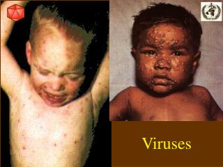
Tobacco mosaic disease stunts growth of tobacco plants and gives their leaves a mosaic coloration • In the late 1800s, researchers hypothesized that a particle smaller than bacteria caused the disease • In 1935, Wendell Stanley confirmed this hypothesis by crystallizing the infectious particle, now known as tobacco mosaic virus (TMV)
Fig. 19-2 RESULTS Extracted sap from tobacco plant with tobacco mosaic disease Passed sap through a porcelain filter known to trap bacteria Rubbed filtered sap on healthy tobacco plants 3 1 2 Healthy plants became infected 4
The Structure of Viruses • Viruses are not cells • Viruses are very small infectious particles consisting of nucleic acid enclosed in a protein coat and, in some cases, a membranous envelope
Viral Genomes • Viral genomes may consist of either • Double- or single-stranded DNA, or • Double- or single-stranded RNA • Depending on its type of nucleic acid, a virus is called a DNA virus or an RNA virus
Capsids and Envelopes • A capsid is the protein shell that encloses the viral genome • Capsids are built from protein subunits called capsomeres • A capsid can have various structures
Fig. 19-3a RNA Capsomere of capsid 18 250 nm 20 nm (a) Tobacco mosaic virus
Fig. 19-3b DNA Capsomere Glycoprotein 70–90 nm (diameter) 50 nm (b) Adenoviruses
Fig. 19-3c Membranous envelope RNA Capsid Glycoproteins 80–200 nm (diameter) 50 nm (c) Influenza viruses
Fig. 19-3d Head DNA Tail sheath Tail fiber 80 225 nm 50 nm (d) Bacteriophage T4
Capsids and Envelopes • Some viruses have membranous envelopes that help them infect hosts • These viral envelopes surround the capsids of influenza viruses and many other viruses found in animals • Viral envelopes, which are derived from the host cell’s membrane, contain a combination of viral and host cell molecules
Bacteriophages • Bacteriophages, also called phages, are viruses that infect bacteria • They have the most complex capsids found among viruses • Phages have an elongated capsid head that encloses their DNA • A protein tail piece attaches the phage to the host and injects the phage DNA inside
Viruses are obligate intracellular parasites, which means they can reproduce only within a host cell • Each virus has a host range, a limited number of host cells that it can infect
General Features of Viral Reproductive Cycles • Once a viral genome has entered a cell, the cell begins to manufacture viral proteins • The virus makes use of host enzymes, ribosomes, tRNAs, amino acids, ATP, and other molecules • Viral nucleic acid molecules and capsomeres spontaneously self-assemble into new viruses
Fig. 19-4 VIRUS Entry and uncoating 1 DNA Capsid Transcription and manufacture of capsid proteins 3 Replication 2 HOST CELL Viral DNA mRNA Capsid proteins Viral DNA Self-assembly of new virus particles and their exit from the cell 4
Reproductive Cycles of Phages • Phages are the best understood of all viruses • Phages have two reproductive mechanisms: the lytic cycle and the lysogenic cycle
The Lytic Cycle of Phages • The lytic cycle is a phage reproductive cycle that culminates in the death of the host cell • The lytic cycle produces new phages and digests the host’s cell wall, releasing the progeny viruses • A phage that reproduces only by the lytic cycle is called a virulent phage • Bacteria have defenses against phages, including restriction enzymes that recognize and cut up certain phage DNA
Fig. 19-5-1 Attachment 1
Fig. 19-5-2 Attachment 1 2 Entry of phage DNA and degradation of host DNA
Fig. 19-5-3 Attachment 1 2 Entry of phage DNA and degradation of host DNA 3 Synthesis of viral genomes and proteins
Fig. 19-5-4 Attachment 1 2 Entry of phage DNA and degradation of host DNA Phage assembly 4 Assembly 3 Synthesis of viral genomes and proteins Head Tail Tail fibers
Fig. 19-5-5 Attachment 1 2 Entry of phage DNA and degradation of host DNA 5 Release Phage assembly 4 Assembly 3 Synthesis of viral genomes and proteins Head Tail Tail fibers
The Lysogenic Cycle of Phages • The lysogenic cycle replicates the phage genome without destroying the host • The viral DNA molecule is incorporated into the host cell’s chromosome • This integrated viral DNA is known as a prophage • Every time the host divides, it copies the phage DNA and passes the copies to daughter cells
Temperate Phages • An environmental signal can trigger the virus genome to exit the bacterial chromosome and switch to the lytic mode • Phages that use both the lytic and lysogenic cycles are called temperate phages
Fig. 19-6 Daughter cell with prophage Phage DNA The phage injects its DNA. Cell divisions produce population of bacteria infected with the prophage. Phage DNA circularizes. Phage Bacterial chromosome Occasionally, a prophage exits the bacterial chromosome, initiating a lytic cycle. Lytic cycle Lysogenic cycle The bacterium reproduces, copying the prophage and transmitting it to daughter cells. The cell lyses, releasing phages. Lytic cycle is induced Lysogenic cycle is entered or Prophage Phage DNA integrates into the bacterial chromosome, becoming a prophage. New phage DNA and proteins are synthesized and assembled into phages.
Reproductive Cycles of Animal Viruses • There are two key variables used to classify viruses that infect animals: • DNA or RNA? • Single-stranded or double-stranded?
Viral Envelopes • Many viruses that infect animals have a membranous envelope • Viral glycoproteins on the envelope bind to specific receptor molecules on the surface of a host cell • Some viral envelopes are formed from the host cell’s plasma membrane as the viral capsids exit
Viral Envelopes • Other viral membranes form from the host’s nuclear envelope and are then replaced by an envelope made from Golgi apparatus membrane
Fig. 19-7 Capsid and viral genome enter the cell Capsid RNA HOST CELL Envelope (with glycoproteins) Viral genome (RNA) Template mRNA Capsid proteins ER Copy of genome (RNA) Glyco- proteins New virus
RNA as Viral Genetic Material • The broadest variety of RNA genomes is found in viruses that infect animals • Retroviruses use reverse transcriptase to copy their RNA genome into DNA • HIV (human immunodeficiency virus) is the retrovirus that causes AIDS (acquired immunodeficiency syndrome)
Fig. 19-8 Viral envelope Glycoprotein Capsid RNA (two identical strands) Reverse transcriptase HIV Membrane of white blood cell HIV HOST CELL Reverse transcriptase Viral RNA RNA-DNA hybrid 0.25 µm DNA HIV entering a cell NUCLEUS Provirus Chromosomal DNA RNA genome for the next viral generation mRNA New virus New HIV leaving a cell
Fig. 19-8a Viral envelope Glycoprotein Capsid RNA (two identical strands) HOST CELL Reverse transcriptase HIV Reverse transcriptase Viral RNA RNA-DNA hybrid DNA NUCLEUS Provirus Chromosomal DNA RNA genome for the next viral generation mRNA New virus
Fig. 19-8b Membrane of white blood cell HIV 0.25 µm HIV entering a cell New HIV leaving a cell
The viral DNA that is integrated into the host genome is called a provirus • Unlike a prophage, a provirus remains a permanent resident of the host cell • The host’s RNA polymerase transcribes the proviral DNA into RNA molecules • The RNA molecules function both as mRNA for synthesis of viral proteins and as genomes for new virus particles released from the cell
Evolution of Viruses • Viruses do not fit our definition of living organisms • Since viruses can reproduce only within cells, they probably evolved as bits of cellular nucleic acid • Candidates for the source of viral genomes are plasmids, circular DNA in bacteria and yeasts, and transposons, small mobile DNA segments • Plasmids, transposons, and viruses are all mobile genetic elements
Mimivirus, a double-stranded DNA virus, is the largest virus yet discovered • There is controversy about whether this virus evolved before or after cells