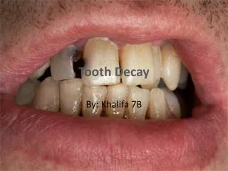
Download
1 / 30
411 likes | 1.23k Views
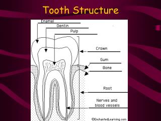
Tooth Development Odontogenesis. Formation of the permanent dentition. during the cap stage the development of the permanent dentition begins – anterior teeth the primordia for these teeth appears as an extension off the developing dental lamina

E N D
Formation of the permanent dentition • during the cap stage the development of the permanent dentition begins – anterior teeth • the primordia for these teeth appears as an extension off the developing dental lamina • penetrates into the mesenchyme lingual to the primary primordium • its site of origin is called the successional dental lamina
Formation of the permanent dentition • these permanent teeth are called succedaneous teeth (anterior teeth and the premolars) • teeth that form with the primary tooth buds (primary predecessors) • permanent molars are non-succedaneous - they are formed by posterior extension of the dental lamina.
Appositional stage • secretion of enamel, dentin and cementum • these tissues are initially secreted as a matrix that is partially calcified – serves as a framework for later calcification • time period varies • multiple inductions occur between the ectodermal tissues of the enamel organ and the mesenchymal tissues of the dental papillae and dental sac
Appositional stage • these inductions are crucial for the production of enamel, dentin and cementum • these interactions are mediated by the basement membrane found in between these ectodermal and mesenchymal tissues • The maturation stage is characterized by the completion of calcification
Ameloblasts • the cells of the IEE assume a more columnar shape or they elongate • differentiate into pre-ameloblasts • this differentiation is characterized by the repolarization of these PAs – movement of the nucleus away from the basement membrane • this repolarization is critical to the differentiation of the PAs
Ameloblasts • continued differentiation and maturation results in the formation of ameloblasts • the pre-ABs induce the cells of the dental papilla to differentiate also
odontoblasts • differentiation by the mesenchyme of the dental papilla • occurs after differentiation of pre-ABs begins • results because the pre-ABs induce differentiation of the mesenchymal cells also • also undergo repolarization – mirror image of the pre-ABs (see Figure 6-12 and 6-13) • after differentiation – the ODs then start dentinogenesis
odontoblasts • begin to deposit predentin on the side of their basement membrane – forms a layer immediately below the BM and above the cells (figure 6-13) • therefore dentin formation begins before enamel synthesis • explains why dentin is thicker than enamel
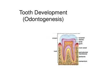
Formation of hard tissues • At 1 the epithelium is separated from the dental papilla by an acellular zone. • At 2 the cells of the inner dental epithelium have elongated, and the acellular zone begins to be eliminated as odontoblasts differentiate from ectomesenchymal cells in the tooth pulp.
Formation of hard tissues • At 3 the odontoblasts retreat toward the center of the pulp, leaving behind formed dentin. • At 4 the cells of the inner dental epithelium, now ameloblasts, begin to migrate outward and leave behind formed enamel. • before dentin forms – cells of the EO receive blood supply from vessels of the dental lamina
Formation of hard tissues • as dentin forms, it cuts of this papillary source of blood/nutrients • this causes a drastic reduction in the amount of nutrients that reach the EO • but the ABs require extensive nutrients to form enamel – stellate reticulum collapses and invagination of the OEE – this brings in blood supply from peripheral vessels found outside the tooth
Formation of hard tissues • after OD differentiation and the initiation of dentinogenesis – the BM between the pre-ABs and ODs disintegrates • this allows direct contact between the pre-ABs and ODs – results in the completion of pre-AB differentiation to mature ABs
Formation of hard tissues • after OD differentiation and the initiation of dentinogenesis – the BM between the pre-ABs and ODs disintegrates • this allows direct contact between the pre-Abs • ABs then begin amelogenesis – apposition of enamel matrix • replaces the disintegrating BM • each ameloblast forms a tapered portion that faces the disintegrating BM - called a tome or Tome’s process
Formation of hard tissues • upon contact of the enamel matrix and dentin – the disintegrating BM begins to mineralize – forms the dentinoenamel junction or DEJ • the ODs form cellular process as they retreat toward the dental papilla (figure 6-14) that penetrate the forming predentin = dentinal tubules • mineralization of the developing dentin and enamel is distinct for each type of tissue • the cell bodies of the ODs remain in the pulp tissue
Formation of hard tissues • the cell bodies of the ABs participate in tooth eruption and will disappear shortly after • and ODs – results in the completion of pre-AB differentiation to mature ABs
Timetable for tooth development • Entire primary dentition initiated between 6 and 8 weeks of embryonic development. • Successional permanent teeth initiated between 20th week in utero and 10th month after birth • permanent molars between 20th week in utero (first molar) and 5th year of life (third molar)
ROOT FORMATION • takes place as the crown is completely shaped and the tooth begins to erupt • therefore the tooth forms from the “top down” – i.e. crown to root • root formation is through the formation of a cervical loop • the CL is the most cervical portion of the enamel/dental organ – two layers consisting of IEE and OEE
ROOT FORMATION • the CL begins to grow down into the dental sac – elongates and moves away from the dental papilla • it forms a Hertwig's root sheath • rim of the sheath = epithelial diaphragm – encloses the developing primary apical foramen • also grows down to encompass all but the basal portion of the pulp
ROOT FORMATION • this sheath shapes the root and induces dentin formation in the root area by the ODs of the dental papilla • ensures that it is continuous with the crown dentin • this sheath lacks the stellate reticulum and stratum intermedium • is capable of differentiating into ODs BUT NOT ABs
ROOT FORMATION • In the case of mandibular teeth the forming front is in a stationary position to the lower border of the mandible • That means that the crown is moving coronally with surrounding tissues and that is a part of the tooth eruption
Multirooted teeth • anterior teeth, premolars and molars all begin as a single root – root trunk • root of the posterior teeth divides from the trunk into the correct number of root branches • differential growth of the H. root sheath results in the division of the root trunk into two or three roots
Multirooted teeth • The root sheath (as a collar hanging from the enamel organ) forms tongues that grow to each other to form many epithelial tubes and secondary apical foramina
Root Dentin • The root of the tooth is composed by dentin and cementum • dentin forms when the outer cells of the dental papilla are induced to differentiation into ODs • similar to what occurs at the crown area • the ODs then begin dentinogenesis and secrete predentin • after dentin formation – the basal membrane disintegrates along with the Hertwig’s sheath
Cementum and Pulp formation • Cementogenesis in the root area also occurs upon degradation of the H. root sheath • the degradation allows contact of the dental sac cells with the dentin surface – induces the formation of cementoblast cells • the CBs cover the root dentin and undergo cementogenesis – laying down cementoid • the CBs do NOT leave cellular processes within the cementum but many CBs become entrapped in the forming cementum
Cementum and Pulp formation • these entrapped CBs are called cementocytes • only upon mineralization of the cementoid can it be called cementum • the region of contact between cementum and root dentin = dentinocemental junction or DCJ • while the cementum is forming - the central cells of the dental papilla form the pulp
Periodontal ligament • the surrounding tissues of the tooth also develop as the crown and root form • the mesenchyme of the dental sac condenses to form the periodontal ligament adjacent to the new cementum • involves synthesis of collagen and bundling into fibers
Periodontal ligament • ends of these fibers insert into the outer layer of cementum and surrounding alveolar bone • the cells of the disintegrating H. root sheath develop into discrete islands of epithelial cells to become epithelial rests of Malassez
Periodontal ligament • The epithelial rests of Malassez become located in the mature periodontal ligament • no known function • they can be identified in the periodontal ligament and are responsible for the development of radicular cysts.