Download
1 / 18
230 likes | 607 Views
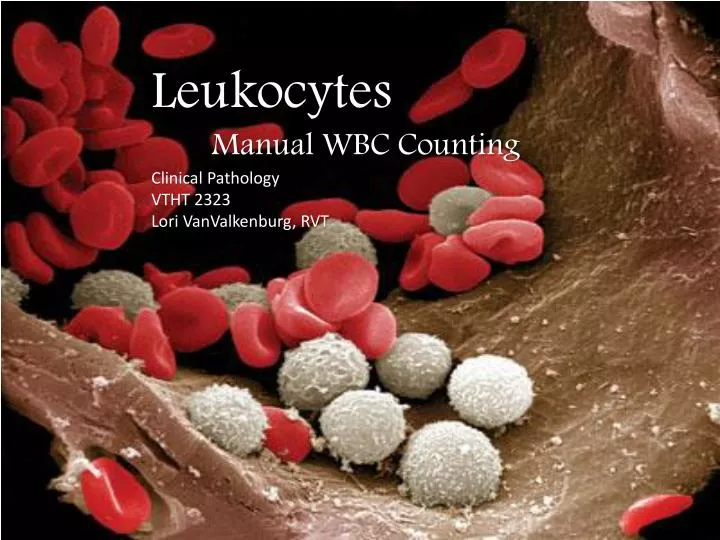
Leukocytes Manual WBC Counting Clinical Pathology VTHT 2323 Lori VanValkenburg , RVT. White Blood Cell Evaluation. Total White Blood Cell Count is the total number of leukocytes in a volume of blood, expressed as thousands/µl may be made manually or with automated cell counters

E N D
Leukocytes • Manual WBC Counting • Clinical Pathology • VTHT 2323 • Lori VanValkenburg, RVT
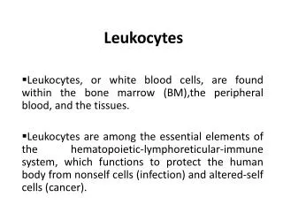
White Blood Cell Evaluation • Total White Blood Cell Count is the total number of leukocytes in a volume of blood, expressed as thousands/µl • may be made manually or with automated cell counters • Manual WBC Count involves utilization of Neubauerhemocytomer and Unopette® dilution system • Automated WBC counters use a variety of methods including: • Impedance • QBC (quantitative buffy coat) • Flow cytometry
Manual Total WBC Count • Unopette system: • 3% Acetic acid diluent lyses cytoplasmic membranes, thereby eliminating RBCs and platelets, leaving behind only nucleated particles. • NeubauerHemocytometer • Counting chamber: An etched grid, consisting of 9 primary squares (0.1 µL per square), used to mark the boundaries for the counting procedure.
Unopette Procedure • Puncture the diaphragm in the neck of the diluent reservoir with the tip of the capillary shield on the capillary pipette.
Unopette Procedure • Remove the protective plastic shield from the capillary pipette. Holding the capillary pipette slightly above the horizontal, touch the tip to the blood source. The pipette will fill by capillary action. When blood reaches the end of the capillary bore in the neck of the pipette, filling is complete and will stop automatically.
Unopette Procedure 3. Use a kimwipe towipe any blood off the outside of the capillary tube, making sure that no blood is removed from inside the capillary pipette 4. With one hand, gently squeeze the reservoir to force some air out, but do not expel any diluent. Maintain pressure on the reservoir. 5. With the index finger or your other hand, cover the upper opening of the capillary overflow chamber and seat the capillary pipette holder in the reservoir neck.
Unopette Procedure 6. Release pressure on the reservoir and remove your finger from the overflow chamber opening. Suction will draw the blood into the diluent in the reservoir. Squeeze the reservoir gently two or three times to rinse the capillary tube, forcing diluent into but not out of the overflow chamber, releasing pressure each time to return diluent to the reservoir. Close the upper opening with your index finger and invert the unit several times to mix the blood sample and the diluent.
Unopette Procedure • Begin timing for 10 minutes • Place the coverglass on the hemocytometer counting chamber, making sure coverglass is clean and free of grease. (Fingerprints must be completely removed.) • Immediately prior to cell counting, mix again by gentle inversion, taking care to cover the upper opening of the overflow chamber with your index finger.
Unopette Procedure • Remove the pipette from the reservoir. Squeeze the reservoir and reseat the pipette in the reverse position, releasing pressure to draw any fluid in the capillary tube into the reservoir. • Invert and fill the capillary pipette by gentle pressure on the reservoir and discard the first 3 drops. • Load (charge) the counting chamber of the hemocytometer by gently squeezing the reservoir while touching the tip of the pipette against the edge of the coverglass and the surface of the counting chamber. A properly loaded counting chamber should have a thin, even film of fluid under the coverglass.
Charging the Hemocytometer • Allow 3 minutes for cells to settle. If fluid flows into the grooves (moats) at the edges of the chamber or if air bubbles are seen in the field, the chamber is flooded and, yes, must be cleaned with distilled water, dried with lens tissue, and reloaded. If the chamber is underloaded, carefully add additional fluid until properly loaded.
Locating the Grid • Carefully place the loaded hemocytometer onto a moistened paper towel (to keep it from drying out) and wait an additional 5-10 minutes for cells to settle completely. • Place the hemocytometer on the microscope. Use the low-power lens to locate the five small fields (1, 2, 3, 4, and 5 in diagram on next slide) in the large center square bounded by the double or triple lines. This is called the “super square” (it is divided into 400 tiny squares) These smaller squares form the grid where RBCs are typically counted.
Each field is composed of 16 small squares. To count the cells in each field, start in the upper left small square and follow the pattern indicated by the arrow in field B. Count all of the cells within each square, including cells touching the lines at the top and on the left. • Do not count any cells that touch the lines on the right or at the bottom. Using the high-power objective (and low light), locate fields A, B, C, and D. Count the WBCs in each of these four corner fields of the hemocytometer chamber.
NeubauerHemocytometer • Hemocytometers are counting chambers used to determine the number of cells per microliter of blood. • The parallel and perpendicular lines that are etched into the two identical sets of fine grids are called “Neubauer rulings”. • The area of each grid is designed to hold a precise amount of sample (0.9 µL) • Each of the 9 squares on the Neubauer grid holds 0.1 µL of sample. • Knowing the number of cells in set parts of the grid and the amount of sample in that area is the basis for calculating the number of WBCs per microliter of blood.
Total WBC Equation The number of cells counted is multiplied by dilution and volume factors to determine total WBC count (in µL) • Add together the number of WBCs observed in each of the 4 large squares then add 9% (to account for 0.9 µL volume of chamber) . Multiply total by 100 (dilution factor) to get total WBC count. Example: Observed 80 WBCs 80 + 7.2 = 87.2 x 100 = 8720/ µL
Accuracy • Both chambers of hemocytometer must be loaded for accurate results. • Counting both sides and comparing results also serve to check accuracy because the number of cells on one side should closely approximate the number of cells on the other side. • An uneven distribution is indicated when there is a variation of more than 15 calls between any of the 4 squares counted. In this case, the count must be discarded.
Possible Sources of Error • Not wiping off excess blood from outside of capillary tube after filling with sample • “Wicking” blood from inside capillary tube when wiping off excess blood from outside of tube • Blood left in capillary tube or accidentally expelled from top of pipette during mixing • Overfilling or underfilling the chamber of the hemocytometer • Be patient. “It will take practice and may require multiple attempts to completely and accurately fill the hemacytometer chamber” (CTVT) • Not allowing cells to settle completely prior to counting • Not adjusting WBC count in presence of NRBCs
Corrected WBC Count • The WBC count, by any method, is a count of nuclei or total nucleated cell count. If nucleated red cells are circulating in blood, they will be included whether the count is done by manual methods or by automated analyzers, falsely elevating the WBC count. • The number of NRBCs per 100 leukocytes is recorded during the differential leukocyte count. If more than 10 NRBCs are counted, then a correction must be made as follows: 100 / 100 + Number of NRBCs x WBCs = Corrected WBC count Example: If 15 NRBCs are counted during the 100-cell differential count, and the initial WBC count is 30,000/µL, the corrected count is calculated as follows: 100 / 100 + 15 x 30,000 = 26,087 WBCs
Normal Total WBC count • Canine: 6,000 – 17,000 /µL • Feline: 5,500 – 19,500 /µL