Download
1 / 21
210 likes | 361 Views
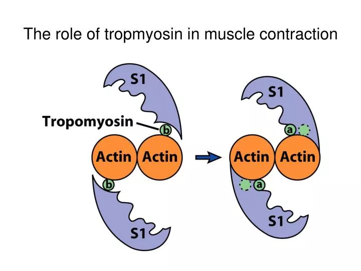
The role of tropmyosin in muscle contraction. 9.7 Nonmuscle motility (1). Actin-binding proteins affect the localized assembly or disassembly of the actin filaments. The roles of actin-binding proteins. Nonmuscle motility (2). Actin-binding proteins (continued)

E N D
9.7 Nonmuscle motility (1) • Actin-binding proteins affect the localized assembly or disassembly of the actin filaments.
Nonmuscle motility (2) • Actin-binding proteins (continued) • Nucleating proteins – provide a template for adding actin monomers. (Arp2/3 complex) • Monomer-sequestering proteins – bind to actin-ATP monomers and prevent them from polymerizing. (thymosin ß4) • End-blocking (capping) proteins – regulate the length of actin filaments. • Monomer-polymerizing proteins –promote the growth of actin filaments. (profilin)
Nonmuscle motility (3) • Actin-binding proteins (continued) • Actin filament depolymerizing proteins – bind actin-ADP subunits for rapid turnover of actin filaments. Example: cofilin • Cross-linking proteins – alter the three-dimensional organization of actin filaments. Examples: vilin, fimbrin
Nonmuscle motility (4) • Filament-severing proteins – shorten filaments and decrease cytoplasmic viscosity. Example: gelsolin • Membrane-binding proteins – link contractile proteins to plasma membrane.
Nonmuscle motility (5) • Examples of Nonmuscle Motility and Contractility • Actin polymerization as a force-generating mechanism • Responsible for some types of motility such as cytoplasmic streaming in Listeria
Nonmuscle motility (6) • Examples of nonmuscle motility and contractility • Cell Locomotion • Cells lacking cilia or flagella move by crawling over a substrate.
Nonmuscle motility (7) • Cell locomotion (continued) • Cells that crawl over a substratum display a repetitive sequence of events.
Nonmuscle motility (8) • Cells that Crawl over the Substratum • Cultured cells crawl by forming a protrusion called a lamellipodium. • Force generation in lamellipodia occurs by adding actin monomers to filaments, prividing temporary anchorage for the cell.
Nonmuscle motility (9) • Axonal Outgrowth • The bulk of the axon shows little evidence of motile activity. • The tip of the axon (growth cone) shows several types of locomotor protrusions: • Microspikes – point outward to the edge of the lamellipodium. • Filopodia – elongations that extend and retract during motile activity.
Nonmuscle motility (10) • Axonal outgrowth (continued) • The growth cone explores its environment and elongates its axon. • Lamellipodia and filopodia of growth cone respond to the presence of physical and chemical stimuli.
Nonmuscle motility (11) • Changes in Cell Shape during Embryonic Development • Ectodermal cells elongate and for a neural plate as microtubules become oriented parallel to the cell’s axis. • Change in cell shape produced by contraction of microfilaments. • Curvature of the neural tube causes outer edges to contact one another forming a tube which gives rise to nervous system.