Download
1 / 28
320 likes | 598 Views
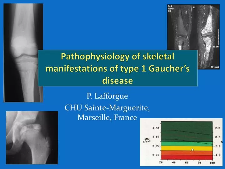
Pathophysiology of skeletal manifestations of type 1 Gaucher’s disease. P. Lafforgue CHU Sainte-Marguerite, Marseille, France. Main cause of pain and disability Most are irreversible Most effective treatment should be prevention . Bone marrow infiltration Pain deformity

E N D
Pathophysiology of skeletal manifestations of type 1 Gaucher’sdisease P. Lafforgue CHU Sainte-Marguerite, Marseille, France
Main cause of pain and disability • Most are irreversible • Most effective treatmentshouldbeprevention.
Bonemarrow infiltration • Pain • deformity • Osteonecroses • Acute bone crises /medullaryinfarcts • Epiphysealosteonecroses • Osteolysis • Osteopenia/osteoporosis • Osteomyelitis • Growth retardation • (multiple myeloma) • Fractures
Deformities • Erlenmeyerflask • 60-80% prevalence • Non specific • Impairment of modeling of dia-metaphysesduringgrowth Faden MA et al. Am J Med Genet A. 2009; 149A: 1334-45
OSTEONECROSES Epiphyseal ON Medullaryinfarcts ≈ 33% of patients • Femoral and humeralheadsmainly • Classicalpresentation … • Chronic pain • disability • Joint replacement • Crises: 20-33% • Rx: 25% • Various locations ++ • Femurs • Tibiae • Pelvicbones, vertebrae … • Osteomyelitis-like crises
Gaucher osteonecroseshallmark • « richmarrow» ON • = sicklecell, Gaucher, leukemias • « hot »: pseudo-osteomyelitis • Bouts of acute crises, atany time • In hematopoïetic/invaded areas: • anywhere, extensive « classical » ON • = idiopathic or secondary (CTC, OH,…) • « cold »: initiallyasymptomatic • Occursimultaneously • In fatty areas : • Epiphyses and metaphyses of long bones
Pathophysiology Vesselslesions Vessels obturation ischeamia Bone/marrownecrosis extrinsicvessels compression
Vascularlesions ? 100 patients • CCL4/MIP-1β • CCL2/MCP-1 • CXCL8/IL-8 +++ • CCL18/PARC • CCL5/RANTES +++ • in Gaucher vs controls • in Gaucher ON vs ON free • Especiallywhen ON occursduring ERT Limits! Treated patients, assessment distant from ON initiation Biological markers of diseaseactivity No evidence of causality Pavlova, Blood Cells Mol Dis 2010
Vascular obturation Micro-emboli of lipidicparticles: corticosteroids dyslipidemia alcoholism Gasbubbles: dysbaric ON thrombosis sicklecell clottingabnormalities
Idiopathic ON • 25 à 30% of ON • 40-50 years-old males • thrombolysis: • Hyperhomocysteinemia • MTFR gene mutation • lipoprotein(a) • tPai Apo B Thrombophilia: S proteindeficiency activated C proteinresistance Prothrombingene mutations Factor V Leiden anti-phospholipid Ab • Inconsistencyacrossstudies • Lack of appropriate control groups • Limits: • High prevalence in general population • High diversity of disorders
capillary adipocyte Bonetrabecule Hématopoieticcells Extrinsicvascular compression extra-vascular components vascularspace Adipocytichypertrophy CTC OH dyslipidemia Medullaryhypertrophy Gaucher’sdisease sicklecell Marrowgasbubbles dysbaric ON intra-medullaryhemorrhages? m. de Gaucher Marrowedema ischaemia: viciouscircle
Riskfactors for ON in GD • Litterature: • correlatedwithmarrow infiltration • splenectomy ++ • male • with ERT • Rodrigue, Clin Orthop 2009; Mistry, BJH 2009 • 56 type 1 GD 24 with / 32 without ON: strong association of ON with: • Youngerageatdiagnnosis • Anyotherbone manifestation • splenectomy • ICGG Registry: 544 GD with ON comparedwith 2008 GD without ON: • Slightly more anemia ( 21,5% vs 11,9%) • Slightly more N270S heterozygoty • Slightly more splenectomy (31% vs 24%), NS • Lanfranchi-Debra 2012, unpublished • Khan A, JBMR 2012 e-pub
Celldeath (osteocytes, marrowcells, adipocytes) collapse Demarcation by a fibrovascularrim, intra-lesionalremodelling Subchondral fracture
Natural course 1) ON are definitive 2) Their volume isfixed 3) The keyeventissubchondral fracture
Natural course 4) Local prognosisdepends on residualmechanicalproperties of the femoralhead. Small ON (<10%) Weightbearing area partiallypreserved X-ray MRI +++ Large ON (>20%) Weightbearing area totallyaffected No symptom Good prognosis Subchondral fracture Deformity Chronic pain
osteolysis • No or few symptoms • Atrisk for fracture
Osteopenia • BMD islower in Gaucher patients Z-score ≈ - 1 SD • However, moderately: adults: T-score < -2.5: 10/57 (18%) • BMD diminution isassociatedwithsplenectomy, hepatomegaly, N370S genotype Javier, Osteoporosis International 2010 Pastores, JBMR 1996 Wenstrup, JBMR 2007
Pathophysiology of osteopenia: GBA1-deficient mouse model • Lower BMD • ↘ stromalcellsproliferation • ↘ OB differentiation • (through PKC inhibition) • Unaffected OC activity Mistry PK et al. PNAS 2010; 107 : 19473-8
Pathophysiology of osteopenia: • Formation: ↘1,2,3,4 • Resorption: ↗1,2 , ↘3 or Nl4 Role of • cytokines: IL-1β, IL-6, IL-10, TNFα ? • MinorlipidLysoGL-1? • Also look for classicalriskfactors 1.Fiore, JBMR 2002; 12 patients 2.Ciana, J InheritMetab Dis 2005; 12 patients 3.Drugan, Blood Cells Mol Dis 2002; 16 patients 4.Van Dussen, J Clin EndocrinolMetab 20011; 40 patients Michelakakis, BiochimBiophys Acta 1996; Allen , QJM 1997; Hollack, Blood Cells Mol Dis 1997
PERIPHERAL FRACTURES • 14-20% • In focal osteolytic areas (pathological fractures ) Stirnemann, Rev Med Int 2006; Javier, Osteoporosis Int 2010
VERTEBRAL FRACTURES • 8-21% of patients T9 T6
VERTEBRAL FRACTURES • Association withlow BMD: logical but no firmevidence • Not associatedwithsplenectomy • Associatedwithoveralskeletalburden Katz, Spine 1993; Stirnemann, Rev Med Int 2006; Javier, Osteoporosis Int 2010 • Khan A, JBMR 2012 e-pub
Pathophysiologyislargelyunknown, but isclearlydriven by medullary infiltration! • Improvementwith ERT or miglustat: • Of pain • Of bone crises number • Of BMD • Few or no new ON: ICCG Registry : follow-up of 2700 Gaucher patients withoutprevalent ON • ERT initiated < 2 yearsafterdiagnosis : ON incidence = 8,1/1000 pts.yrs • ERT initiated > 2 yearsafterdiagnosis : ON incidence = 16,6/1000 pts.yrs • Risk x 2 whenhistory of splenectomy • However complications maystilloccurundertherapy Charrow, Clin Genet 2007 Sims, Clin Genet 2008 Mistry, Br J Haematol 2009 Stiernemann, ArthritisResTher 2010
Open prospective trial • 33 patients (M=43 years) ERT-naivetreatedwithimiglucerase Bone pain BALP ostéocalcin Bone markers FN BMD Lumbar BMD DPyru NTXu Sims et al, Clin Genet 2008
Children • ICGG Registry • 884 chidrenreceiving ERT , 8 yearsfollow-up • height: Z-score -1,4 normal (-0,3) • 90 patients withprevalentbone crises : 0 crises after2 yearstherapy • 440 patients withoutprevalentbone crises 2,5% withsubsequent new crises • BMD: Z-score -0,35 +0,29 SD Fig1, 6 et 7 Andersson et al. Pediatrics 2008 height Newcrises Lumbar BMD