Download
1 / 79
830 likes | 1.08k Views
Advances in Imaging: Echo, CT, CMR. Justin D Pearlman MD ME PhD Director, Dartmouth Advanced Imaging Center. Disclosures. Consultant for: General Electric Picker/Marconi/Phillips Chiron Boehringer-Ingelheim MagnaLab Perfusion=off-label use of contrast.

E N D
Advances in Imaging:Echo, CT, CMR Justin D Pearlman MD ME PhD Director, Dartmouth Advanced Imaging Center
Disclosures • Consultant for: • General Electric • Picker/Marconi/Phillips • Chiron • Boehringer-Ingelheim • MagnaLab • Perfusion=off-label use of contrast
Dartmouth Advanced Imaging Center - Aims High-end imaging capabilities; Bench->Bedside • 1. Realtime CMR • 2. 4D Cardiac CT • 3. 3D Echo, PET • Viability • Myopathy • Microcirculation • Dx • Rx
Equipment Echo MR CT
Echo short axis CMR short axis Images: CMR vs. Echo Cost: $500-$1500 Cost: $400-800
Echo Inject packets of energy waves, pulsed, Receive echoes Scan to collect data to convert to image Tomographic Dynamic Flow signal from phase shift CMR Insert packets of energy waves, pulsed, Receive echoes Scan to collect data to convert to image Tomographic Dynamic Flow signal from phase shift Similarities
Sound Speed 1540 m/s Echo=A-mode (amp-time) Stopped by Metal Bone Air-tissue Views limited by rib window, contact, angle Resolution depends on frequency, beamwidth Bright blood requires contrast Radiowave Speed 299,792,258 m/s Echo=K-mode (amp-spatial freq) Distorted by Metal (No problems with bone, air/tissue) Any view Resolution is adjustable down to 10 microns, limited by noise and acquisition time Bright blood many ways Differences Echo CMR
Basis for MRI B0 Gx,y,z + B1 Mz Mxy + +
Change in Magnetization T1/TR 1-e(-TR/T1) 1-2e(-TI/T1) T1/TI T2/TE e(-TE/T2)
K-space sums 3,2 2,5 = + Sum (3,2) + 0.5 (2,5) 0.5 (3,2) + (2,5) (3,2) + (2,5)
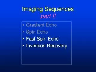
Methods • Magnetization Preparation • Excitation • Spatial Encoding, Echoes • Image Reconstruction Bright Blood TurboGradient Echo, Dark Blood FSEDIR, Fat-suppresive TIR, … (100’s)
Echo Intense Short T1 (if T1-weighted image) Short T2 (if T2-weighted image) Water (if fat suppressed) Fat (if not fat suppressed) Corresponding notions
Fat vs. Fat Suppression: RVD Fat+ vs. Fat- RV RV JDP 2/02
SMART function Pearlman JD et alSerial motion assessment by reference tracking (SMART): application to detection of local functional impact of chronic myocardial ischemia. J Comput Assist Tomogr, 2001. 25(4): p. 558-62
Calcium Scoring • Claims: • Negative score may indicate non-cardiac sources of chest pain • Scores over 1,000 predict coronary event within the next 2-3 years • Positive scores referred for catheterization or stress test • BUT significant disease may have negative score • Positive score may be stable plaque
Elastic Match of Coronaries • Fast CT of mom • Elastic match • contrast • Simulated holography as background, for context
Perfusion-Sensitive Imaging Resting delayed blood arrival predicts ischemia
Rest Delayed Blood Arrival Dark Late Zone Arrived
Space-Time Map We introduced Space- Time Maps to see delay in blood arrival in a single derived image
Table 1: Clinical Characteristics of Study Population Total number Patients N % Prevalence SE Disease vessel 105 38 97 2.69 0.10 CABG 42 10 26 1.08 0.13 Angioplasty 36 22 56 0.92 0.14 Stent 15 11 28 0.38 0.11 Agreement between Rest MRI and Rest / Stress Nuclear Rest MRI vs. rest Thallium / stress MIBI
Scar RV TV septum RA LVOT LV lateral LA MV Delayed Enhancement 62 year old patient with 3-vessel CAD c/o angina at rest.Hx MI 1992, PTCA LAD 1992, CABG 1995.Scintigraphy, MRI : lateral + anteroseptal wall defects
Molecular Imaging bFGF2
Microvascular MRI • Tissue bright • Major vessels visible • Dynamic physiology • Small vessels hidden
Angiogenesis-Sensitive MRI r=.95 No contrast Dark Flash 3D CT Validation Radiology 214:801 ‘00 Acad Rad 4:680 ’97 Nat Med 1:1085 ‘95
Dark Flare PredictsImprovedBlood Arrival From Angiogenesis Baseline 1 Month 2 Months
Dark Flare/Delayed Arrival Combined First Dose-Response for Angiogenesis Rx DA=Demand, CX=Response
Angiogenesis imaging may also help diagnose and treat cancer 34 y.o. woman with a palpable breast mass. Ultrasound negative Mammography negative Collateral Sensitive MRI: fat black, collateral neovascular development flashes; cancer found.
MRI Microscopy in Large Target Limit signal to 1 cm2 Fold-over problem Avoid fold-over Look at bowl of kiwi 40 micron resolution RME = Response- Modulated Excitation No fold-over Fold-over
What to know • Vocabulary • B0, B1, Mz, Mxy, T1, T2, T2*,, , , • TI, TR, TE, , Matrix, FOV, , TD, TW • GE, SE, FISP, HASTE, … • Tilted Tomographic Anatomy • Pathophysiology, Clinical Decisions • Physics, Image Processing
Echo vs. CMR CMR Echo “Both are watching out for the CAT skinner”
Clinical Example • 42 y.o. man with large cell lymphoma • Radiation to chest • Paroxysmal atrial fibrillation • CT: Mediastinal mass ? LA compression
Long Axis 4 Chamber View Echo MRI: mass, effusion
Long Axis 2 Chamber View Echo: ? NL fxn MRI:effusion,mass