Download
1 / 55
590 likes | 796 Views
Basic Immunology 101. Amy Sharma Ph.D. Candidate Uetrecht Laboratory Leslie Dan Faculty of Pharmacy, University of Toronto. Q. Why does your immune system exist?. Immunology Overview. Immune system is like a double edged sword Key players of the immune system

E N D
Basic Immunology 101 Amy Sharma Ph.D. Candidate Uetrecht Laboratory Leslie Dan Faculty of Pharmacy, University of Toronto
Immunology Overview • Immune system is like a double edged sword • Key players of the immune system • Humoral versus Cellular Immunity
I. Cells of the immune system • T-lymphocytes (Thymus derived ‘T’ cells) • Key role in cell-mediated immunity • co-ordinate and regulate immune responses through cytokine activation, antibody stimulation, etc • Constitute ~60-70% of lymphocytes in circulating blood • Many different sub-types • Identified by T-cell receptor
I. Cells of the immune system • B-lymphocytes (Bone marrow derived ‘B’ cells) • Key role in humoral immunity: • produce antibodies against antigens • act as antigen-presenting cells (APCs) • develop into memory B cells after activation by antigen interaction • Constitute ~10-20% of lymphocytes in circulating blood
I. Cells of the immune system • Macrophages (“Big-eaters”) • Key role in immunity in general: • main type of APC (process and present antigen to CD4+ Th-cells) • phagocytose and kill microbes coated by antibody and/or complement • produce cytokines, regulating T and B cell function
I. Cells of the immune system • Dendritic cells (“Potent” APC) • Key role: link between adaptive and cell mediated immunity • process antigen and present peptide fragments to other cells of the immune system goes on to regulate T and B cell responses
I. Cells of the immune system • Natural Killer ‘NK’ cells (“LGL’s”) • Key role in cell-mediated immunity • contain azurophilic granules thus capable of lysing tumor cells, virus infected cells, etc, without previous sensitization • Constitute ~10-15% of lymphocytes in circulating blood
Innate Immunity (cellular immunity) • Mediated by lymphocytes • Does not involve antibodies (antigen non-specific) • Cellular immunity protects the body by: • activating macrophages, NK cells, and cytotoxic T-cells • stimulating cytokine secretion, influencing the function of other immune cells
Humoral Immunity • Mediated by soluble antibody proteins (antigen specific) • Humoral immunity protects the body by: • antigen presentation, discriminating recognition of “non-self” versus “self” • the generation of antibody responses • the development of immune memory
Idiosyncratic Drug ReactionsWhat are They, Why & How Do We Study Them? Amy Sharma Ph.D. Candidate Uetrecht Laboratory Leslie Dan Faculty of Pharmacy, University of Toronto
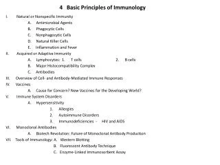
Overview • Adverse Drug Reactions (ADRs) • Idiosyncratic Drug Reactions (IDRs) • Characteristics of IDRs • Proposed Mechanism of IDRs • Drugs Known to Induce IDRs • Studying IDRs • Future Directions
I. Adverse Drug Reactions • The World Health Organization definition: “any noxious, unintended, and undesired effect of a drug, which occurs at doses used in humans for prophylaxis, diagnosis, or therapy” • ADRs are common • 2,216,000 hospitalized patients/year experienced a serious ADR and 106,000/year died from an ADR • Fatal ADRs rank 4th to 6th in leading causes of death in US (Bond CA et al. Pharmacotherapy 2006)
I. Adverse Drug Reactions Adverse drug reactions can be divided into five basic types: • Type A (augmented): • Can be predicted from the pharmacology of the drug • Are typically dose-dependent • Type C (chemical), D (delayed) and E (end of treatment) • Type B: • Cannot be predicted on the basis of the known pharmacology of the drug • Also known as idiosyncratic adverse reactions • Can affect almost any organ system
II. Idiosyncratic Drug Reactions • Rare & unpredictable reactions • Incidence: 1/103 - 1/106 patients • 25% of all ADRs • Still very prevalent because of the number of drugs involved and the number of people taking these drugs • Do not occur in most patients at any dose • No simple dose-response relationship • Effects not related to pharmacological properties of the drug • Can be very severe • most serious ADRs in drug therapy
Bone marrow / Blood cells (aplastic anemia; agranulocytosis) III. Characteristics of IDRs • Organs affected: • Most thought to be immune-mediated • Detected during the late stage of development or when drug is released on to market • May lead to withdrawal • Significant financial burden Liver (cholestatic liver) Skin (mild-severe rash)
III. Risk Factors for IDRs: • Don’t have a good understanding of who will develop IDRs. • Age - Incidence increases with age • Concomitant challenge – increase risk for HIV patients • Ethnic background – Incidence of clozapine-induced agranulocytos is 20% in a Jewish hospital vs. <1% elsewhere • Gender - female >> male
IV. Mechanisms of IDRs If we can understand how drugs induce IDRs we can: • Scan for drugs that have high risk of causing IDRs early in the drug development process, and avoid later losses to both patients and manufacturers • Devise therapy that prevents IDRs in patients (administer concomitant therapy) There is circumstantial evidence that indicates a potential role of reactive metabolites (RMs) in development of IDRs
IV. Step 1: Reactive Metabolite Formation • Drug Metabolism: • Process whereby therapeutically active drugs are converted to a more soluble form (metabolites) and are cleared by renal or biliary excretion • Reactive Metabolites (RMs) and Covalent Binding • During metabolism, usually through P450 oxidation, drugs can form RMs (chemically reactive species) that can covalently bind to endogenous proteins or other macromolecules
Reactive Metabolites • Reactive metabolites are electrophiles or free radicals • Sulfates/sulfonates • Epoxides/arene oxides • Michael Acceptors • Nitroso amines
IV. Where Does Metabolism Occur? Metabolizing enzymes are present in the following organs: Cytochrome P450, Sulphotransferases, Peroxidases White blood cells (macrophages and neutrophils) that become activated to kill bacteria, and do so by releasing oxidants such as H2O2 and HOCl.
IV. Where Does Metabolism Occur? Once formed, reactive metabolites tend to bind to nucleophilic groups on proteins or macromolecules near the site of their formation. Thus, toxicity most often occurs at sites of RM formation, especially if RM is highly reactive! Example – Clozapine: • Clozapine is oxidized to a RM in both the liver and neutrophils. The main toxic effects of clozapine are liver and neutrophil toxicity (hepatotoxicity and agranulocytosis).
IV. Step 2: Immune Response • Basic paradigm in Immunology • To discriminate against pathogens, the immune system learns to recognize self from non-self. In this way, autoimmunity is avoided and immune responses are mounted against foreign invaders. • Hapten Hypothesis • Once drug is covalently bound to a host protein it forms a novel antigen known as the hapten-carrier complex. Host immune system then perceives the modified endogenous protein as foreign, and mounts an immune response against it.
IV. Hapten Hypothesis Detailed Step 1 – Reactive Metabolite Formation Step 2 – T-cell activation and Initiation of an Immune Response IDR
1. Reaction takes several weeks to develop 2. Once the drug is removed, reaction clears quickly 3. On re-exposure the time to onset is shorter than on first exposure 4. In some reactions anti-hapten antibodies or antibodies against self-tissues are found (e.g., in patients with halothane-induced hepatitis anti-hapten antibodies have been found) IV. IDR Characteristics that Indicate Immune Involvement Not all IDRs have these characteristics
V. Clinical Evidence in Support of Hapten Hypothesis • Penicillin-induced anaphylaxis • Aminopyrine-induced agranulocytosis • Halothane-induced hepatitis
-lactam ring Benzylpenicillin V. Penicillin-Induced Anaphylaxis • Covalent binding due to spontaneous ring opening • IgE antibodies were detected in patients with anaphylactic reaction • Re-exposure can be life-threatening
V. Aminopyrine-Induced Agranulocytosis Myeloperoxidase/H2O2/Cl- Dication intermediate • Associated with a high risk of agranulocytosis (~1%) • Reactive dication formed by neutrophil-derived hypochlorous acid could be responsible for the IDR • Onset of symptoms (fever, sore throat and infections) in 1 week - 1 month • Drug-specific Abs • Re-challenge results in rapid drop in neutrophil count as well as their bone marrow precursors
Protein F O Br F O F P4502E1 Cl F C C C l C C F F C C NH F H F F Halothane RM covalently bound to protein Trifluoroacetyl Chloride V. Halothane-Induced Hepatitis • Halothane is oxidized by P450 to form trifluroacetyl chloride, which can bind to proteins • 20% of patients develop asymptomatic elevation of liver transaminases (AST, ALT) • leads to the development of hepatitis • hepatitis rarely occurs on first exposure, which suggests that sensitization is required • Serum of affected patients contain antibodies against native hepatic proteins as well as trifluoroacetylated proteins (hapten-carrier complex)
V. Drugs Known to Cause IDRs Felbamate antiepileptic Nevirapine HIV drug (NNRTI) D-Penicillamine anti-rheumatic Clozapine antipsychotic Carbamazepine anticonvulsant
Idiosyncratic reactions: Aplastic Anemia and Liver Toxicity Reactive Metabolite: Yes Protein Binding: Probable Animal Model: No V. Felbamate Phenylacrolein (Michael Acceptor)
Idiosyncratic reaction: Severe Skin Rash, Liver Toxicity Reactive Metabolite: quinone methide Protein Binding: Yes; epidermis Animal Model: Yes; Skin rash in the female Brown Norway rat V. Nevirapine Quinone Methide
V. Nevirapine skin rash Human skin in response to NVP treatment Female rat skin in response to NVP treatment
Idiosyncratic reaction: Autoimmunity, lupus Reactive Metabolite: None, parent drug can bind to proteins through the thiol group Protein Binding: Yes Animal Model: Yes; Autoimmunity in the male Brown Norway rat V. D-Penicillamine Forming mixed disulfides
Idiosyncratic reaction: Agranulocytosis, Liver Toxicity, Cardiac Toxicity Reactive Metabolite: Yes Protein Binding: Yes Animal Model: No V. Clozapine Nitrenium Ion
Idiosyncratic reaction: Anticonvulsant hypersensitivity syndrome (fever, rash, multi-organ involvement etc.) Reactive Metabolite: Yes Protein Binding: Yes Animal Model: No V. Carbamazepine Iminoquinone
VI. Methods Ideally want to illustrate each step for each drug: 1. Metabolism 2. Reactive Metabolite Formation 3.Protein Binding in Target Tissue(s) 4.Immunogenicity of Hapten 5.Immune Response IDR
VI. Step 1: Metabolism – Microsomes Mince liver in sucrose buffer Homogenize Excise liver Centrifuge 100,000 x g Centrifuge 10,000 x g cytosol S9 fraction microsomes nuclei, cell membrane mitochondria
Analyze reaction mixture by HPLC or LC/MS Confirm identity of products with NMR (require pure metabolite) Incubate at 37C a) Buffer (physiological conditions, salt/ pH7.4) b) Microsomes c) Drug d) NADPH generating system (NADP+, G6PD, G6P) VI. Step 1: Metabolism – Microsomes
Upper solution is placed on top of a ficoll solution (density gradient) Obtain (human / rat) blood Sediment RBCs with dextran Plasma, WBCs Ficoll RBCs Centrifuge ~1000 rpm Pour off upper layers, use remainingneutrophils lymphocytes neutrophils VI. Step 1: Metabolism - Neutrophils
Analyze reaction mixture by HPLC or LC/MS Confirm identity of products with NMR (require pure metabolite) Incubate at 37C a) Buffer (physiological conditions, salt/ pH7.4) b) Neutrophils c) Drug d) Neutrophil activator (PMA) VI. Step 1: Metabolism - Neutrophils
VI. Step 2: RM Formation • Complete same experiments as when looking at metabolism but with an additional step • Reactive metabolite may be so reactive that it is not detected on the HPLC chromatogram • Must add GSH or NAC to the reaction mixture to trap the reactive metabolite in a stable form that can be detected by HPLC and later identified by LC/MS and NMR
VI. Step 3: Protein Binding in Target Tissues • Require an antibody that recognizes the reactive metabolite (the hapten) • Must prepare antigen by linking the reactive metabolite to an immunogenic carrier protein e.g., KLH • Immunize rabbits with this antigen • Sera obtained from the blood of these rabbits is polyclonal, and contains antibodies againstthehapten
In Vivo: Isolate and homogenize target tissues Administer drug to rats or mice VI. Step 3 Cont’d • Complete in vivo and in vitro studies • in vitro studies are similar to metabolism studies • in vivo studies involve administering the drug to animals (rats or mice)
VI. Step 3 Cont’d Take tissues from either in vitro or in vivo experiment and perform Western blot analysis to detect covalent binding of reactive metabolites to proteins: • Run the protein sample on an SDS polyacrylamide gel • Transfer separated proteins from gel to nitrocellulose membrane • Blot membrane with an antibody against the HAPTEN • Visualize antibody binding with a detection system; presence of covalent adducts will thus be elucidated