Download
1 / 63
660 likes | 961 Views
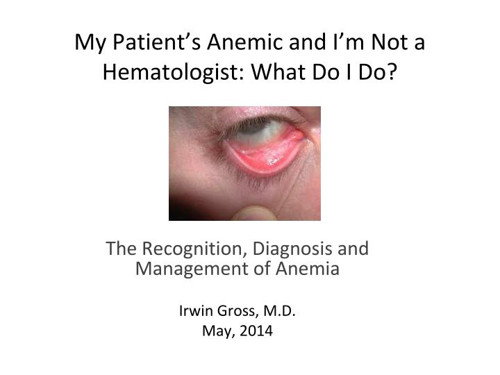
Iron Poor “Tired Blood”. My Patient’s Anemic and I’m Not a Hematologist: What Do I Do?. The Recognition, Diagnosis and Management of Anemia Irwin Gross, M.D. May, 2014. No Disclosures. Prevalence of Anemia. Estimated prevalence of anemia in U.S. is 3.5 million

E N D
Iron Poor “Tired Blood” My Patient’s Anemic and I’m Not a Hematologist: What Do I Do? The Recognition, Diagnosis and Management of Anemia Irwin Gross, M.D. May, 2014
Prevalence of Anemia • Estimated prevalence of anemia in U.S. is 3.5 million • Almost certainly an under-estimate • Previously undiagnosed anemia is common in elective surgical patients • Most common underlying causes include: • Iron deficiency • Vitamin B12 deficiency • Chronic kidney disease • Other chronic inflammatory diseases • Folate deficiency (uncommon in U.S.) • Unexplained Anemia of the Elderly (UAE)
Iron Deficiency in Pediatrics • Prevalence • 9% of 1-3 year olds are iron deficient with 3% IDA • Rates decrease with age until adolescence, then increase when 15% of females are iron deficient • Increased prevalence with decrease in socioeconomic status • Increased prevalence with obesity • Poor intake of iron-rich foods • Obesity is a chronic inflammatory state • Perinatal risk factors • Maternal iron deficiency • Prematurity • Insufficient dietary iron intake in early infancy
Iron Deficiency and Neurocognitive Development • Possible relationship between ID/IDA and neurobehavioral development • Recent RCT in LBW (2,000 – 2,500 gm) neonates • 285 infants randomized to 0, 1, or 2 mg/kg/day of enteric iron supplements from 6 weeks to 6 months of age • Assessed at 3.5 years along with 95 normal BW controls • Results • No difference in IQ between LBW groups or LBW vs. controls • Significant difference in behavioral problems based on Child Behavior Checklist (CBCL) • Scores above the U.S. cutoff: 12.7%, 2.9%, and 2.7% respectively for 0, 1, and 2-mg groups compared with 3.2% in controls Pediatrics 2013; 131:47-55
Gender disparity in anemia prevalence Kassenbaum et al. Blood 2013 doi:10.1182/blood-2013-06-508325
Global prevalence of anemia approximately 30% ! Kassenbaum et al. Blood 2013 doi:10.1182/blood-2013-06-508325
Anemia – It’s an Epidemic! Anemia is a common complication of common diseases • 30-60% of patients with RA have anemia • 30-80% of patients with IBD have anemia • 30-50% of patients with CHF have anemia • 20-40% of diabetics without overt renal failure have anemia • 40-60% of patients with chronic kidney disease have anemia All of these are related to iron absorption and metabolism
Age, Anemia and Iron Deficiency • 17% of adults over the age of 65 have iron deficiency • Of those with iron deficiency anemia, only 50% normalized their hemoglobin with oral iron therapy • 35% of adults over the age of 65 have unexplained anemia (defined as hemoglobin less than 12 g/dl Blood Cells Mol Dis. 2011;46(2):159
Why We Should Care (More) About Anemia? • The value of recognizing and treating anemia goes beyond reducing the risk of transfusion • Effective clinical management of anemia improves patient outcomes in CHF, CKD, IBD, rheumatoid diseases, etc. • Anemia may an indicator of an undiagnosed underlying disease process, e.g. iron deficiency suggesting occult malignancy
Preoperative Anemia and Postoperative Outcomes in Non-cardiac Surgery • ACS NISQIP Database study: 227,425 subjects, > 18 years; major non-cardiac surgery, excluding trauma • 30% of patients were anemic • Patients with even mild anemia (hgb 10 - 12g/dl in women; 10 - 13 g/dl in men) experienced: • Higher 30 day adjusted mortality • Increased morbidity including cardiac, respiratory, urinary tract, wound events, sepsis and thromboembolism • Perioperative transfusion also independently associated with increased morbidity and mortality • Treatment of preoperative anemia should be strongly considered • Transfusion is…”the least favorable option” Musallam Lancet 2011;378:1396-407
Impact of Anemia in Patients with AMI Undergoing PCI: Analysis from the CADILLAC Trial JACC 2004;44(3): 547-553
Iron Deficiency in CHF: An International Pooled Analysis Iron Deficiency (with and without anemia) in 40% of NYHA I and 60% of NYHA IV CHF patients
Etiology of Anemia • Common causes of anemia • Iron deficiency • Chronic blood loss • Nutritional deficiency: most common nutritional deficiency worldwide and the most common cause of anemia • Chronic kidney disease (often with iron deficiency) • Chronic heart failure (often with iron deficiency) • Hypothyroidism • Anemia of inflammation, acute or chronic • Most common anemia in hospitalized patients • Often co-exists with iron deficiency • Unexplained anemia of the elderly (UAE)
Etiology of Anemia • Less common causes of anemia • Disorders of hemoglobin synthesis, e.g. thalassemia • Autoimmune hemolytic anemia • Anemia associated with non-hematologic malignancy • Myelodysplasia and other primary clonal abnormalities of the marrow
Unexplained Anemia of the Elderly • Some anemic patients have unexplained anemia of the elderly (UAE) • Usually mild (hgb 11-12 g/dL) but associated with poor outcomes • Approximately 50% have elevated IL-6, CRP, or other markers of inflammation • Low EPO levels • Really a diagnosis of exclusion • If macrocytic, think B12 deficiency, folate deficiency or MDS
Laboratory Evaluation of Anemia • Basic evaluation • CBC • Include reticulocyte count • Iron, iron binding capacity, ferritin • Vitamin B12 • Creatinine • Consider as second tier tests • TSH • Folate • DAT • Haptoglobin • LDH
Laboratory Evaluation of Anemia • First review the clinical history, then review the CBC: • Review the red cell indices (will discuss in a moment) • Look for abnormalities in the platelet count • Thrombocytosis: iron deficiency, inflammation or myeloproliferative disease • Thrombocytopenia: many causes, including medications and increases risk of bleeding • Look for abnormalities in number and/or type of white cells • Inflammatory process • Infection • Leukemia, lymphoma, myelodysplasia
Laboratory Evaluation of Anemia • Hemoglobin, hematocrit and red cell count • Hemoglobin reference range: • 14.0 – 17.0 g/dL (male) • 12.0 – 15.0 g/dL (female) – Is Hgb of 12 g/dl truly “normal”? • Hematocrit reference range: • 42 – 51% (male) • 36 – 45% (female) • Red cell count reference range • 4.7 – 6.0 M/µL (male) • 3.9- 5.5 M/µL (female)
Laboratory Evaluation of Anemia • Hemoglobin, hematocrit and red cell count • For purposes of pre-operative anemia screening, we use a hemoglobin less than 13 g/dL as “anemia” for all patients • Hemoglobin < 13 g/dL is “inflection” point for increased risk of transfusion with high blood loss procedures • Recognize that some women with hemoglobin of 12-13 g/dL are normal and that some men with hgb between 13-14 g/dl are not • Hemoglobin is directly measured and is the more “accurate” measure if Hgb and Hct don’t “agree” • Estimated Hct is usually (Hgb value) x 3 • Anemia with elevated red cell count is seen in thalassemia minor
Laboratory Evaluation of AnemiaRed Cell Indices • MCH and MCHC: Mean corpuscular hemoglobin (pg) and corpuscular hemoglobin concentration g/dL (in a given volume of blood) • Of minimal value • MCV: Mean Cell Volume • Most useful of the red cell indices • Measured value in femtoliters (fl) • Reference range 80-100 fl • A primary tool for categorizing anemia • < 80 fl = microcytic • >100 fl = macrocytic
Laboratory Evaluation of AnemiaRed Cell Indices • MCV: Mean Cell Volume • Microcytic anemia (< 80 fl): • Iron deficiency • Thalassemia or other disorders of heme synthesis • Normocytic anemia (80-100 fl) • Anemia of chronic inflammation usually normocytic • Anemia associated with CKD (may be macrocytic) • Anemia associated with hypothyroidism (may be macrocytic) • Iron deficiency anemia and B12 deficiency anemia may also be normocytic
Laboratory Evaluation of AnemiaRed Cell Indices • MCV: Mean Cell Volume • Macrocytic anemia (>100 fl) • Anemia due to B12 or folate deficiency usually macrocytic (but may be normocytic) • Hemolytic anemias are often macrocytic • Anemia secondary to alcohol • Anemia secondary to chronic liver disease • If macrocytosisis associated with ovalocytosis and/or other cytopenias consider possibility of a myelodysplastic syndrome (MDS)
Laboratory Evaluation of AnemiaReticulocytes • Reticulocyte count: • Decreasedin hypoproliferative anemia relative to the degree of anemia (e.g. nutritional deficiency anemia, CKD, ACI, etc.) • “Normal” reticulocyte count in an anemic patient suggests an inadequate marrow response • Increased in hemolytic anemia or blood loss with marrow response and in response to initiation of effective therapy • Best measure is absolute reticulocyte count, not percentage, reticulocyte index or reticulocyte production index • Reference range: 25,000 – 85,000 / microliter
Laboratory Evaluation of AnemiaBeyond the CBC • Labs to evaluate iron status: • Serum iron • Reference range 50 – 150 µg/dL • Decreased in iron deficiency and anemia of acute or chronic inflammation • Diurnal variation with highest levels in the morning • As much as a 30% variation within day and between day due to changes in marrow iron uptake, iron absorption, storage iron outflow, etc. • Fasting sample!
Laboratory Evaluation of AnemiaBeyond the CBC • Labs to evaluate iron status: • Transferrin • The principal plasma protein for transport of iron • Total Iron Binding Capacity (TIBC) is an indirect measure of transferrin concentration and is often used interchangeably with transferrin • TIBC expressed in µg/dL (Reference range: 260-475 µg/dL) • Reference range: 200-400 mg/dL • Increased in iron depletion states • Decreased in inflammatory states including anemia of chronic inflammation and in malnutrition, liver disease, malignancy
Laboratory Evaluation of Anemia Beyond the CBC • Labs to evaluate iron status: • Transferrin saturation (TSAT) • Reference range: 20-45% saturation • < 20% consistent with iron deficiency or functional iron deficiency • > 45% suggests iron overload • Transient increase above 45% typical with I.V. iron Rx • Percent saturation reflects iron available for erythropoiesis • Lower limit of current laboratory reference ranges is too low (in many labs it is < 12-15%)
Laboratory Evaluation of Anemia Beyond the CBC • Labs to evaluate iron status: • Ferritin • Reference range: > 100 ng/mL (upper limit varies with age) • MOST labs use a lower limit of 12-20 ng/mL for females and 30-45 ng/ml for males; this is too low • < 100 ng/ml suggests decreased iron stores • HMW protein consisting of about 20% iron by weight • Found in all cells but especially hepatocytes and macrophages serving as an iron reserve
Laboratory Evaluation of AnemiaBeyond the CBC • Labs to evaluate iron status: • Ferritin (cont’d) • A small amount found in plasma and reflects iron stores in normal individuals • Acute phase reactant - often markedly increased in acute illness regardless of iron status, so increased ferritin does not rule out iron deficiency • Low ferritin is best single laboratory indicator of iron depletion, but a normal or elevated ferritin does NOT rule out iron deficiency
Studies that Assist in Evaluating Iron Deficiency Anemia and Anemia of inflammation
Laboratory Evaluation of AnemiaBeyond the CBC Other labs: • Vitamin B12: • Reference range 200-900 pg/mL • Some patients with B12 between 200-300 pg/mL will also be deficient • If B12 is < 300, consider treatment with B12 or further evaluation with serum methyl malonic acid • If < 200, treat • Up to 15-20% of patients with B12 deficiency will be normocytic, not macrocytic
Laboratory Evaluation of AnemiaBeyond the CBC Other labs: • Folate • Reference range: 5-40 ng/mL serum; 280-900 ng/mL red cell folate • I usually don’t test for folate deficiency • Isolated folate deficiency is uncommon, i.e. there is a low prevalence of folate deficiency in the U.S. • I add oral folate to any treatment regimen for anemia • If you measure folate (e.g. in a patient with macrocytic anemia and a normal B12) • Red cell folate better than serum folate but requires a different blood sample • Serum folate is acceptable IF a fasting sample
Laboratory Evaluation of AnemiaBeyond the CBC Other labs: • Creatinine • Chronic kidney disease (CKD) is a common cause of anemia – 40% of Stage 4 and 5 CKD • Anemia secondary to CKD is, in part, a diagnosis of exclusion • A glomerular filtration rate estimated from the creatinine (eGFR) can be used to qualify patients for anemia treatment with an ESA • CRP • Increased CRP indicates inflammation
Laboratory Evaluation of AnemiaBeyond the CBC Other labs: • Tests for hemolysis • Direct antiglobulin test (positive) • LDH (elevated) • Haptoglobin (decreased) • Bilirubin (elevated, especially indirect) • Reticulocyte count (elevated) • Thyroid Stimulating Hormone (TSH) • Hypothyroidism may be a cause of anemia, usually normocytic
Key Points! • Hemoglobin is directly measured and is the more accurate measure if hgb and hct don’t agree • For purposes of pre-operative anemia management we use a hgb of < 13g/dl as “anemia” (male or female)since that is the inflection point for increased risk of transfusion • Use the absolute reticulocyte count to determine if the anemia is hypoproliferative (thousands/microliter) • Ferritin is an acute phase reactant and does not reflect iron stores in a patient with inflammation
Key Points! • The lower end of published reference ranges for ferritin and transferrin saturation are too low • Ferritin less than 100 ng/ml suggests depleted iron stores • Transferrin saturation less than 20% suggests decreased iron available to the “erythron” for red cell production • Increased ferritin and decreased transferrin saturation is consistent with so-called functional iron deficiency, previously “anemia of chronic disease” or “anemia of chronic inflammation”
Key Points! • Many patients with iron deficient erythropoiesis are not anemic, but still symptomatic from their iron deficiency • Many of the symptoms of iron deficiency anemia are due to iron deficiency, not the anemia • Iron deficiency anemia and B12 deficiency anemia can both be normocytic • Iron deficiency anemia is usually microcytic and B12 deficiency anemia is usually macrocytic, but… • The MCV is normal in 15-20% of patients with B12 deficiency • Many patients with iron deficiency or functional iron deficiency have a normal MCV (not microcytic)
When you’ve done all those tests and you still don’t have a diagnosis • Still no etiology, consider a referral to a hematologist for bone marrow examination and further evaluation • Underlying malignancy • Myelodysplasia • Plasma cell dyscrasia
Iron Deficiency, Functional iron Deficiency and Iron Deficiency Anemia
Iron Deficiency Anemia • Most common cause in U.S. is blood loss • GI lesion • Menstrual or abnormal uterine bleeding • Hematuria • Decreased iron absorption another major cause • Atrophic gastritis, H. pylori, celiac disease, use of PPIs and H2 -antagonists • Gastric bypass surgery • Vegetarian diet • Crohn’s disease, celiac disease, giardiasis • Chronic use of NSAIDs • In pediatrics, cow’s milk at early age
How Much Iron? • Elemental iron requirement: 0.27 mg/day for term infants increasing to about 7 mg/day at 7-12 months • 1-2 mg daily in healthy adults • Daily iron requirement • Replace iron lost from epithelial cell sloughing (skin, GU, GI) • Iron for increased blood volume • Iron for increased tissue mass • Iron for stores
Maximum Daily Iron Absorption Maximum daily enteric iron absorption: 5-7 mg
Hepcidin • Most important protein for regulating iron absorption, release and transport • Hepcidin is decreased by anemia, hypoxia, iron deficiency • Decreased hepcidin results in increased iron absorption • Hepcidin is increased by iron loading AND systemic inflammatory response • Systemic inflammation results in iron restricted erythropoiesis (i.e. F.I.D.) due to up-regulation of hepcidin • This is known as “hepcidin blockade”
Iron Absorption and Hemostasis Hepcidin, Inflammation and Iron Metabolism Lysosomal degradation IV Iron IV iron leads to translational and post-translational up - regulation of ferroportin via IRP + IRP’s 1,2 J Am SocNephrol 18: 394-400, 2007 Blood 2005 106: 3979-3984
“Functional” Iron Deficiency • Chronically ill patients often have functional iron deficiency (FID) despite normal or increased ferritin • Include: • Post-operative and trauma patients • Patients with IBD, RA and related diseases, or other chronic inflammatory process • Cardiac patients • Some overlap with Unexplained Anemia of the Elderly (UAE) • Patients with FID: • Are characterized by low iron, low iron binding capacity, and low iron saturation • May have normal or increased ferritin
What all this means about iron absorption and bioavailability • If there is inflammation: • Enteric iron absorption is significantly impaired • Release of iron from storage is significantly impaired • Ferritin may be increased • Transferrin saturation will be decreased • Erythroid (and other) cells will be deprived of adequate iron and become functionally iron deficient • Anemia results • FID is the most common cause of “anemia of chronic disease”
Clinical Manifestations of ID • Signs and symptoms are non-specific • Impaired myocardial function • Increased mortality? • Impaired immune function • Weakness, fatigue, lethargy • Headache • Irritability • Exercise intolerance • Cognitive dysfunction • Pica, especially for ice (pagophagia) • Poor feeding • Restless leg syndrome