Download
1 / 3
30 likes | 36 Views
The clinical and laboratory studies states that diabetic retinopathy is the major cause of permanent blindness among the aged personalities. The problem with this disease is that there is no cure for it and the only thing we can do is to detect the disease as soon as possible in order to prevent further loss of vision. In this system we propose a CNN approach for diagnosing DR from retinal images and classifying the stages of the disease .The classification is done based on the haemorrhages, micro aneurysms present in the retinal image. We train this network using a high end graphics processor unit GPU using kaggle data set and the disease classification is done, hence we can identify the the disease and prevent the further loss of vision. N Rahul | Roy Eluvathingal | Sanith Jayan K | Mr. Anil Antony "Diabetic Retinopathy Detection using Neural Networking" Published in International Journal of Trend in Scientific Research and Development (ijtsrd), ISSN: 2456-6470, Volume-4 | Issue-4 , June 2020, URL: https://www.ijtsrd.com/papers/ijtsrd31487.pdf Paper Url :https://www.ijtsrd.com/engineering/computer-engineering/31487/diabetic-retinopathy-detection-using-neural-networking/n-rahul<br>

E N D
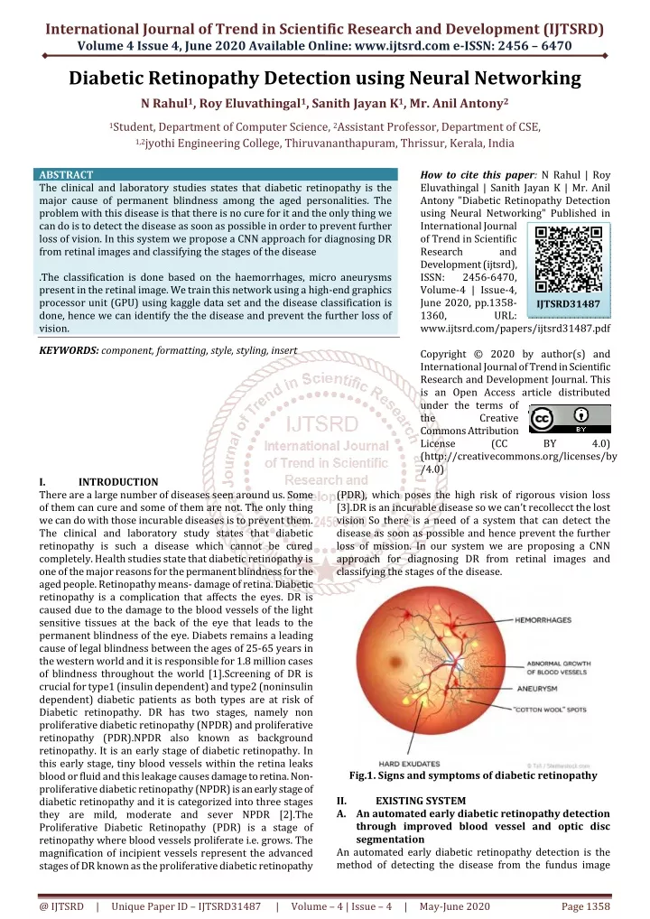
International Journal of Trend in Scientific Research and Development (IJTSRD) Volume 4 Issue 4, June 2020 Available Online: www.ijtsrd.com e-ISSN: 2456 – 6470 Diabetic Retinopathy Detection using Neural Networking N Rahul1, Roy Eluvathingal1, Sanith Jayan K1, Mr. Anil Antony2 1Student, Department of Computer Science, 2Assistant Professor, Department of CSE, 1,2jyothi Engineering College, Thiruvananthapuram, Thrissur, Kerala, India ABSTRACT The clinical and laboratory studies states that diabetic retinopathy is the major cause of permanent blindness among the aged personalities. The problem with this disease is that there is no cure for it and the only thing we can do is to detect the disease as soon as possible in order to prevent further loss of vision. In this system we propose a CNN approach for diagnosing DR from retinal images and classifying the stages of the disease .The classification is done based on the haemorrhages, micro aneurysms present in the retinal image. We train this network using a high-end graphics processor unit (GPU) using kaggle data set and the disease classification is done, hence we can identify the the disease and prevent the further loss of vision. KEYWORDS: component, formatting, style, styling, insert How to cite this paper: N Rahul | Roy Eluvathingal | Sanith Jayan K | Mr. Anil Antony "Diabetic Retinopathy Detection using Neural Networking" Published in International Journal of Trend in Scientific Research and Development (ijtsrd), ISSN: 2456-6470, Volume-4 | Issue-4, June 2020, pp.1358- 1360, URL: www.ijtsrd.com/papers/ijtsrd31487.pdf Copyright © 2020 by author(s) and International Journal of Trend in Scientific Research and Development Journal. This is an Open Access article distributed under the terms of the Creative Commons Attribution License (CC (http://creativecommons.org/licenses/by /4.0) IJTSRD31487 BY 4.0) I. There are a large number of diseases seen around us. Some of them can cure and some of them are not. The only thing we can do with those incurable diseases is to prevent them. The clinical and laboratory study states that diabetic retinopathy is such a disease which cannot be cured completely. Health studies state that diabetic retinopathy is one of the major reasons for the permanent blindness for the aged people. Retinopathy means- damage of retina. Diabetic retinopathy is a complication that affects the eyes. DR is caused due to the damage to the blood vessels of the light sensitive tissues at the back of the eye that leads to the permanent blindness of the eye. Diabets remains a leading cause of legal blindness between the ages of 25-65 years in the western world and it is responsible for 1.8 million cases of blindness throughout the world [1].Screening of DR is crucial for type1 (insulin dependent) and type2 (noninsulin dependent) diabetic patients as both types are at risk of Diabetic retinopathy. DR has two stages, namely non proliferative diabetic retinopathy (NPDR) and proliferative retinopathy (PDR).NPDR also known as background retinopathy. It is an early stage of diabetic retinopathy. In this early stage, tiny blood vessels within the retina leaks blood or fluid and this leakage causes damage to retina. Non- proliferative diabetic retinopathy (NPDR) is an early stage of diabetic retinopathy and it is categorized into three stages they are mild, moderate and sever NPDR [2].The Proliferative Diabetic Retinopathy (PDR) is a stage of retinopathy where blood vessels proliferate i.e. grows. The magnification of incipient vessels represent the advanced stages of DR known as the proliferative diabetic retinopathy INTRODUCTION (PDR), which poses the high risk of rigorous vision loss [3].DR is an incurable disease so we can’t recollecct the lost vision So there is a need of a system that can detect the disease as soon as possible and hence prevent the further loss of mission. In our system we are proposing a CNN approach for diagnosing DR from retinal images and classifying the stages of the disease. Fig.1. Signs and symptoms of diabetic retinopathy II. A.An automated early diabetic retinopathy detection through improved blood vessel and optic disc segmentation An automated early diabetic retinopathy detection is the method of detecting the disease from the fundus image EXISTING SYSTEM @ IJTSRD | Unique Paper ID – IJTSRD31487 | Volume – 4 | Issue – 4 | May-June 2020 Page 1358
International Journal of Trend in Scientific Research and Development (IJTSRD) @ www.ijtsrd.com eISSN: 2456-6470 through the improved segmentation strategies for optic and blood vessels. It proposes improved techniques like micro aneurysms as well as haemorrhages detection which helps in finding the disease as soon as possible. The idea we are taken from this pa-per is the improved blood vessel and optic disc segmentation. The aim of mass screening programs for diabetic retinopathy is to detect and diagnose the disorder earlier than it causes loss of vision. This system consist of mainly 5 stages. The first stage is pre-processing. In this stage the pre-processing of the eye fundus image is done for detecting the disease. The main objective of preprocessing technique is to increase the detection probability of disease by visual assessment and computer aided segmentation of retinal images. After this stage detection blood vessels are done from the retinal image. Foreground and background classification of image is done during this stage. The third stage is optic disc segmentation where the complete partition of the fundus image is done. After the optic disc segmentation the fovea of the eye is detected. Fovea is the part of the eye where large number of blood vessels are seen. This is appeared as a very dark color in the fundus image of eye.All the pathological structures, namely vascular tree, optic disc and fovea which may cause false positive, are removed from the fundus image [4]. The remaining patho-logical structures like micro aneurysm and haemorrhages are extracted from colour fundus image. The last stage is feature extraction of micoaneurysm and hemorrhage. After extracting micro aneurysms and hemorrhages from fundus image, feature selection takes place in order to acquire relevant information from the given image. Based on this feature extraction the stages of the diabetic retinopathy is identified and the result is shown by the system. B.Hardware based analysis on automated early detection of Diabetic-Retinopathy Hardware based detection of diabetic retinopathy isn’t an easy problem to solve. With an increase in bit depths and image sizes software based detection techniques are becoming less useful. Real time systems need to be quick in processing and be able to take in a large amount of information. DSP based systems help to ensure that only relevant information is passed to the human analyst. Fundus images are the input to the system. These images need to be preprocessed before it is passed to the segmentation stage. Process involves in this stage includes color space conversion, zero padding of image edges, median filtering, contrast stretching and windowed based adaptive histogram equalization with overlap mean [5]. C.Diabetic Retinopathy: Present and Past A lot of work has been done in this field and there are various ways for detecting DR like detecting blood vessels, various lesions such as micro aneurysms, exudates, hemorrhages etc. A change in shape and size of blood vessels is a good indicator of detecting DR. Presence of various lesions helps in detecting diabetic retinopathy. Thus various researches have been bifurcated in two ways as of automating blood vessels segmentation. First method is segmentation of retinal blood vessels. It means to separate the blood vasculature of retina in fundus images from its background. But this is a difficult task because of low contrast in fundus imaging, variable size vessels, presence of various pathologies as micro aneurysms, hard exudates, hemorrhages etc. Vessel segmentation can be achieved by supervised, unsupervised or deep neural net-works. Supervised methods make use of training set which is manually processed and segmented by ophthalmologists [6].The second method is identification of various lesions. Here various preprocessing steps such as colour normalization, contrast enhancement along with fuzzy C- means clustering have been applied for image segmentation. III. METHOD AND STRUCTURE A.Structure of Neural Network We used the technique called Transfer learning to implement the neural network. The base of our network was made up of an already trained network called ResNet50 and we fine tuned it by adding our own layers. A sequential model was created with ResNet50 as first layer and fine tuning was donw by adding a Global Average Pooling layer along with a combination of dense and dropout layers. All dense layers use ReLu as activation function except for the last layer which uses softmax as the activation function. Dropout layers were used to reduce over fitting. The model is capable of detecting 5 stages of Diabetic retinopathy ie; No DR, Mild DR, Moderate DR, Severe DR and Proliferative DR. Fig.2. Fine tuned Neural Network structure B.Dataset, Software and Hardware The dataset was obtained from kaggle (APTOS 2019 Blindness Detection) which consisted 3,662 images with @ IJTSRD | Unique Paper ID – IJTSRD31487 | Volume – 4 | Issue – 4 | May-June 2020 Page 1359
International Journal of Trend in Scientific Research and Development (IJTSRD) @ www.ijtsrd.com eISSN: 2456-6470 labels 0, 1, 2, 3, 4 describing levels of diabetic retinopathy. We resized the image so that training is easier without losing much details from the image. We used Kaggle Notebooks which is a free cloud computational jupyter environment (Similar to Google Colab). We used Tensor Flow, Keras along with several other packages for developing the model. Open CV was used for image processing. C.Preprocessing The dataset contained images of patients of different age, ethnicity and the lightning was different in each fundus imaging. Colour normalisation was done to bring all the fundus images into a common lighting. Open CV was used for colour normalisation. The images was of high resolution so it was reduced to 220x220 pixels so that training will be faster without losing much details of the image. Auto cropping was also used to remove unwanted black areas from the image. D.Training Out of the 3,662, 2929 images were used for training and 733 images were used for validation. 10 epoch were done to train the network. Early stopping was implemented with validation loss as minimum set as the parameter and patience as 5. So if a validation loss from an epoch is set as minimum and the next 5 epochs doesn’t produce a even lesser validation loss, the the training will be stopped. This was done to save time, fasten the training process and reduce over fitting. The best model obtained was saved. IV. RESULT Out of 3,662 images, 733 were saved for validation purpose. The validation process took 170 seconds to complete. The classification is as follows 0-No DR, 1-Mild DR, 2-Moderate DR, 3-Severe DR, 4-Proliferative DR. Accuracy is defined as the amount of patients with correct classification. The accuracy obtained for the best model after validation was 78 percent. Fig.4. Confusion matrix V. Diabetic retinopathy (DR) is the leading eye disease in our world. It causes permanent blindness. With this system we are going to detect the disease from its early stage hence we can control the further loss of vision. The lost vision cannot be recollected. The only thing we can do is to detect the disease from its starting stage and prevent the further loss of vision. Here in this system we are taking the input image from the patients (retinal image) and after processing the image the system will extract the features from the image and classifies the stages of the disease. With the help of the neural network technology the system will generate very efficient and accurate result. This system will help the people to detect the disease from its early stages and prevent the further loss of vision. REFERENCES [1]Kumar, Shailesh, and Basant Kumar. ”Diabetic retinopathy detection by extracting area and number of microaneurysm from colour fundus image.” 2018 5th International Conference on Signal Processing and Integrated Networks (SPIN). IEEE, 2018. CONCLUSION [2]Rajput, Yogesh M., et al. ”Detection of non-proliferative diabetic retinopathy lesions using wavelet and classification using K-means clustering.” 2015 International Conference on Communication Networks (ICCN). IEEE, 2015. [3]Kasurde, Snehal Dilip, and S. N. Randive. ”An automatic detection of proliferative diabetic retinopathy.” 2015 International Conference on Energy Systems and Applications. IEEE, 2015. 271–350. [4]Kumar, Shailesh, et al.”An automated early diabetic retinopathy detec-tion through improved blood vessel and optic disc segmentation.” Optics & Laser Technology 121 (2020): 105815. [5]Datta, N. S., et al.”Hardware based analysis on automated early detection of Diabetic-Retinopathy.” Procedia Technology 4 (2012): 256-260. [6]Gupta, retinopathy: Present and past.” Procedia computer science 132 (2018): 1432-1440. Ankita, and Rita Chhikara. ”Diabetic Fig.3. Model loss and Model accuracy @ IJTSRD | Unique Paper ID – IJTSRD31487 | Volume – 4 | Issue – 4 | May-June 2020 Page 1360