Download
1 / 8
80 likes | 84 Views
Antifungal potential of u00c3u0178 citronellol was studied on C. albicans, a commensal that causes both superficial and systemic infection in immunocompromised humans. This non toxic natural compound displayed a minimum inhibitory concentration MIC of 200 u00b5-g mL and was fungicidal at 350 u00b5-g mL. On YPD agar, it formed inhibition zones ZOI of 16.2 mm, 18.15 mm and 19.25 mm respectively, at MIC, 2MIC and 3MIC while fluconazole FLC formed ZOI of 20.43 mm at its MIC disc diffusion assay . No growth was observed above 2MIC on solid media spot assay and = MIC in liquid YPD media. Hydrolytic enzyme secretion decreased in the presence of u00c3u0178 citronellol and was concentration dependent. At MIC, the reduction in phospholipase activity 54.08 was greater than the reduction in proteinase activity 40.6 . There was concentration dependent decrease in total ergo sterol content by 19 , 40 , 91 and 100 , respectively at MIC 8, MIC 4, MIC 2 and MIC values of u00c3u0178 citronellol. FLC at MIC showed an inhibition of only 64 . Biofilm formation reduced by 71.13 at MIC. u00c3u0178 citronellol, hence has immense antifungal potential and significantly inhibits growth, ergosterol levels, hydrolytic enzyme secretion, adhesion and biofilm formation in C. albicans at MIC and sub MIC values. Sumit Kumar Rastogi | Yamini Sharma | Nikhat Manzoor "Effect of u0392--Citronellol on Hydrolytic Enzyme Secretion, Ergo sterol Biosynthesis and Bio film Formation in C. Albicans" Published in International Journal of Trend in Scientific Research and Development (ijtsrd), ISSN: 2456-6470, Volume-2 | Issue-5 , August 2018, URL: https://www.ijtsrd.com/papers/ijtsrd18305.pdf Paper URL: https://www.ijtsrd.com/biological-science/microbiology/18305/effect-of-u03b2-citronellol-on-hydrolytic-enzyme-secretion-ergo-sterol-biosynthesis-and-bio-film-formation-in-c-albicans/sumit-kumar-rastogi<br>

E N D
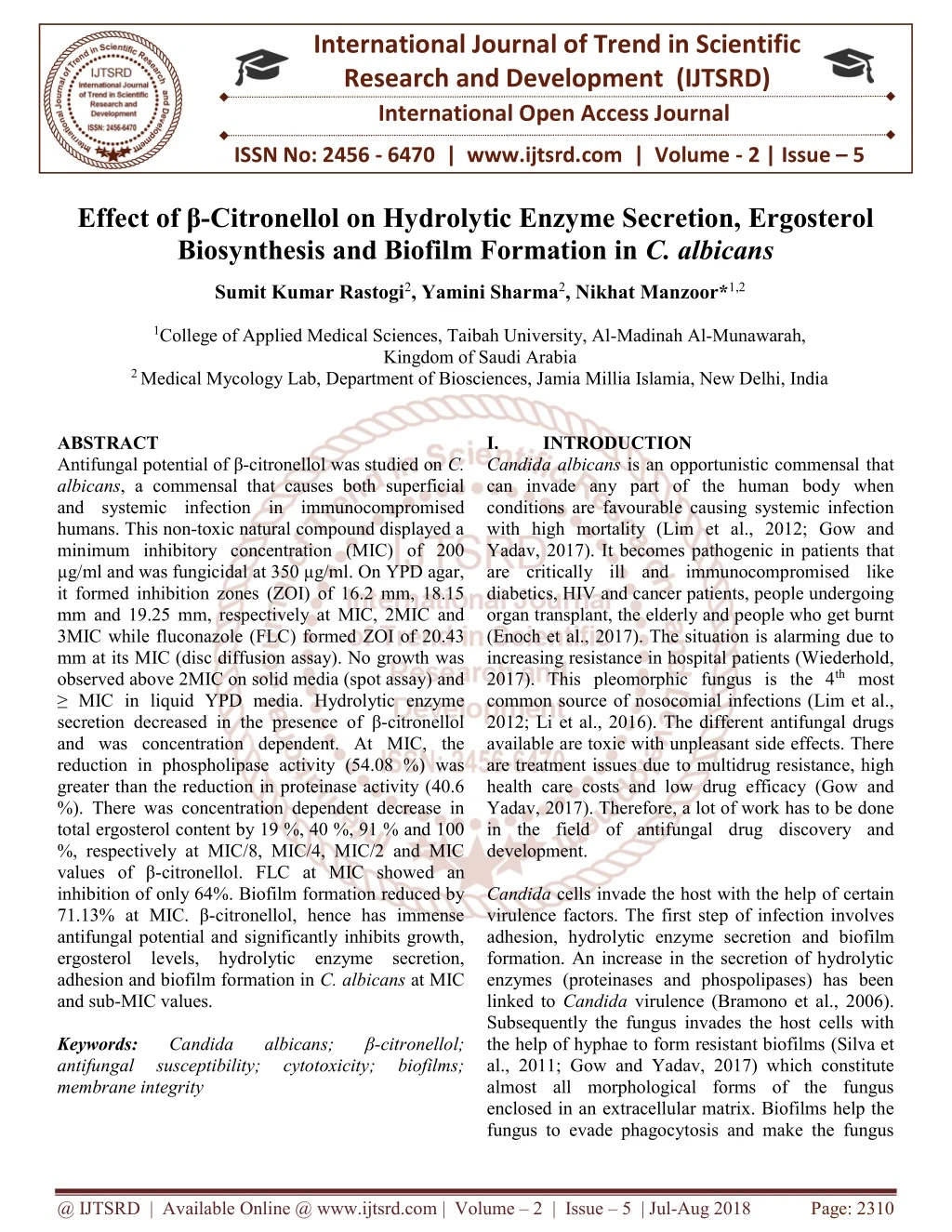
International Journal of Trend in Scientific Research and Development (IJTSRD) International Open Access Journal ISSN No: 2456 - 6470 | www.ijtsrd.com | Volume - 2 | Issue – 5 Effect of β-Citronellol on Hydrolytic Enzyme Secretion, Ergosterol Biosynthesis and Biofilm Formation in C. albicans Sumit Kumar Rastogi2, Yamini Sharma2, Nikhat Manzoor*1,2 1College of Applied Medical Sciences, Taibah University, Al-Madinah Al-Munawarah, Kingdom of Saudi Arabia 2 Medical Mycology Lab, Department of Biosciences, Jamia Millia Islamia, New Delhi, India ABSTRACT Antifungal potential of β-citronellol was studied on C. albicans, a commensal that causes both superficial and systemic infection in immunocompromised humans. This non-toxic natural compound displayed a minimum inhibitory concentration (MIC) of 200 µg/ml and was fungicidal at 350 µg/ml. On YPD agar, it formed inhibition zones (ZOI) of 16.2 mm, 18.15 mm and 19.25 mm, respectively at MIC, 2MIC and 3MIC while fluconazole (FLC) formed ZOI of 20.43 mm at its MIC (disc diffusion assay). No growth was observed above 2MIC on solid media (spot assay) and ≥ MIC in liquid YPD media. Hydrolytic enzyme secretion decreased in the presence of β-citronellol and was concentration dependent. At MIC, the reduction in phospholipase activity (54.08 %) was greater than the reduction in proteinase activity (40.6 %). There was concentration dependent decrease in total ergosterol content by 19 %, 40 %, 91 % and 100 %, respectively at MIC/8, MIC/4, MIC/2 and MIC values of β-citronellol. FLC at MIC showed an inhibition of only 64%. Biofilm formation reduced by 71.13% at MIC. β-citronellol, hence has immense antifungal potential and significantly inhibits growth, ergosterol levels, hydrolytic enzyme secretion, adhesion and biofilm formation in C. albicans at MIC and sub-MIC values. Keywords: Candida antifungal susceptibility; membrane integrity I. Candida albicans is an opportunistic commensal that can invade any part of the human body when conditions are favourable causing systemic infection with high mortality (Lim et al., 2012; Gow and Yadav, 2017). It becomes pathogenic in patients that are critically ill and immunocompromised like diabetics, HIV and cancer patients, people undergoing organ transplant, the elderly and people who get burnt (Enoch et al., 2017). The situation is alarming due to increasing resistance in hospital patients (Wiederhold, 2017). This pleomorphic fungus is the 4th most common source of nosocomial infections (Lim et al., 2012; Li et al., 2016). The different antifungal drugs available are toxic with unpleasant side effects. There are treatment issues due to multidrug resistance, high health care costs and low drug efficacy (Gow and Yadav, 2017). Therefore, a lot of work has to be done in the field of antifungal drug discovery and development. Candida cells invade the host with the help of certain virulence factors. The first step of infection involves adhesion, hydrolytic enzyme secretion and biofilm formation. An increase in the secretion of hydrolytic enzymes (proteinases and phospolipases) has been linked to Candida virulence (Bramono et al., 2006). Subsequently the fungus invades the host cells with the help of hyphae to form resistant biofilms (Silva et al., 2011; Gow and Yadav, 2017) which constitute almost all morphological forms of the fungus enclosed in an extracellular matrix. Biofilms help the fungus to evade phagocytosis and make the fungus INTRODUCTION albicans; cytotoxicity; β-citronellol; biofilms; @ IJTSRD | Available Online @ www.ijtsrd.com | Volume – 2 | Issue – 5 | Jul-Aug 2018 Page: 2310
International Journal of Trend in Scientific Research and Development (IJTSRD) ISSN: 2456-6470 less sensitive to conventional antifungals (Sardi et al., 2013; Lohse et al., 2018). β-citronellol, a natural possesses, anti-inflammatory (Brito et al., 2012), insect repellent (Semmler et al., 2014), larvicidal (Hierro et al., 2004), and anti-bacterial (Lopez- Romero et al., 2015) properties. Like geraniol, it is a major component of rose oil and a number of other plant essential oils. Chemically, β-citronellol (dihydrogeraniol) is related to geraniol whose anti- Candida activity and mode of action was studied recently (Sharma et al., 2016). Although its antifungal potential has been reported (Pereira Fde et al., 2015) its mode of action is not clearly discussed. In the present study, we observed the effect of varying concentrations of β-citronellol on growth pattern, secretion of proteinases and phospholipases secretion, membrane ergosterol content and biofilm formation in C. albicans ATCC 10261. II. MATERIAL AND METHODS C. albicans ATCC 10261 was maintained on YPD media constituting yeast extract, peptone, dextrose, in a percentage ratio of 1:2:2 supplemented with 2.5% agar at 4°C. All chemicals were of analytical grade purchased from Merck components, β-citronellol and fluconazole (FLC) were purchased from Sigma Aldrich (Germany). 2.1. Antifungal drug susceptibility assays Broth dilution method (CLSI, 2008) was used to determine the minimum inhibitory concentrations (MIC) defined as the least possible concentration that causes 90% decrease in absorbance in comparison to that of the control (without test compound). After getting the MIC value, 15 µL aliquots were removed from tubes that show absolutely no growth along with the last tube showing growth. These were sub- cultured on YPD agar plates and incubated at 35 ºC until growth was visible in the control samples. The minimum fungicidal concentration or MFC value was determined as the minimum concentration of the test compound for which there was no visible growth (Samber et al., 2015). Results were calculated as mean of the two separate experiments with three different values. To study antifungal susceptibility to β-citronellol (Ansari et al., 2014) by spot assay, Candida cells were grown overnight in YPD media at 37 ºC, and suspended in 0.9% NaCl to achieve an optical density of 0.1 at 600 nm. Aliquots (5 µL) of five-fold serially diluted cultures were pipetted on YPD agar plates in the absence (control) and presence of MIC and sub- MIC values of β-citronellol. After 48 h of incubation at 30 ºC, growth variations were observed. Candida cells (1x10⁵cells/ml) were inoculated into YPD agar at 40 ºC and poured into 90 mm petriplates to perform disc diffusion assay. Sterile filter discs (4mm) were laden with three different concentrations of test compound (MIC, 2MIC and 3MIC) and placed on agar plates. After 48 h, the average diameter of inhibition zones (ZOI) was measured. A disc impregnated with FLC at its MIC value was used as positive control. Candida cells were sub-cultured at least two times and grown till stationary phase is reached at 35 °C on SDA plates. To study growth pattern, cell culture (A595 = 0.1) was inoculated into fresh media along with β-citronellol at MIC and sub-MIC concentrations in 50 ml total volume. Growth was monitored at 37 °C, 200 rpm and recorded after every 2 h till 48 h. Absorbance was recorded at 595 nm for each concentration using Labo-med Inc. spectrophotometer (USA) and plotted against time in hours. 2.2. WST-1 based cytotoxicity assay The assay was performed as described previously (Khan et al., 2010). Cell culture (1×10⁵ cells/ml) was taken along with β-citronellol (MIC and sub-MIC values) in 96-well plate (final volume ~ 100 µl/well) and incubated for 24 h. WST- 1/CEC dye (10 µL) was added to each well and plates were again incubated at 37 ºC for 2 h with shaking. The reaction was stopped by adding 10 µL of 1% SDS. WST-1 salt was reduced to red coloured formazan by cellular dehydrogenases (Tsukatani et al., 2008), the absorbance of which was recorded at 450 nm using a micro plate Reader (BIORAD iMark, US) (reference was set at 655 nm). Experiment was repeated thrice and cytotoxicity was calculated using the following equation: % Cytotoxicity = [(cell control – cell with test compound)/(cell control)] ×100 2.3. Secretion of hydrolytic enzymes For assessing the activity of proteinases and phospholipases, Candida cells were first inoculated into 5 ml YPD media and incubated for 18 h at 37 °C (Khan et al., 2014). Subsequently, cells were separated from culture media, washed twice and re- suspended in 0.9% NaCl. (MacFarland 0.5 index) were exposed to desired cyclic monoterpenoid, (India). The media Cell suspensions @ IJTSRD | Available Online @ www.ijtsrd.com | Volume – 2 | Issue – 5 | Jul-Aug 2018 Page: 2311
International Journal of Trend in Scientific Research and Development (IJTSRD) ISSN: 2456-6470 concentrations of β-citronellol (1/8 MIC, ¼ MIC, ½ MIC and MIC). In case of proteinase assay, small aliquots (2 µl) were placed at equidistant points on agar plates (2% agar, 0.2 g BSA, yeast nitrogen base, 20 g glucose, and distilled water to a final volume of 1000 ml) while in case of phospholipase assay, aliquots were pipetted on agar peptone media (2 % agar, 10 g peptone, 30 g glucose, 57.3 g NaCl, 0.55 g CaCl2, and distilled water to a final volume of 900 ml) enriched with 10 % (v/v) egg yolk emulsion (HiMedia). Plates were incubated at 37 °C till noticeable growth (2-4 days). Enzyme secretory activity was estimated degradation/precipitation zones formed and calculated in terms of Pz values which is the ratio of the colony diameter to the colony diameter plus diameter of the zone of degradation/precipitation (Price et al., 1982). 2.4. Adhesion and biofilm formation These experiments were performed according to Ramage et al., 2002 with modifications. An aliquot of 100 µl cell suspension (1x10⁷cells/ml) in RPMI 1640 media was placed into each well of a sterile 96-well microtiter plate. β-citronellol was added to each well at desired concentrations except control. Plates were incubated at 37ºC with gentle shaking to allow the cells to adhere to plate walls. After 90 min, non- adherent cells were washed out of each well with PBS (150µl). To see effect on biofilm formation, after the initial 1 h adhesion period, freshly prepared media (RPMI 1640) containing desired concentrations of β- citronellol were added to the wells containing adherent cells. The plates were then incubated for 24 h at 37°C. Estimation was done by semi-quantitative XTT reduction assay. A saturated solution of XTT (in PBS) was mixed with menadione-acetone solution (electron mediator). Pre-formed Candida bio films were washed with PBS first and taken in a 96-well plate with or without β-citronellol. A 100 μl aliquot of XTT was then added to each well and mixed gently. The plates were incubated in dark for 5 h at 37 °C after which absorbance of each sample was recorded at 450 nm using a micro titre plate reader (BIO-RAD, iMark, US). The wells containing PBS only or PBS + XTT were used as blank. The results were expressed as percentage viability. 2.5. Sterol extraction and quantitation Sterol content of treated and untreated Candida cells was evaluated as discussed earlier with slight changes (Sharma et al., 2016). Briefly, 50 ml of SD broth (along with varying concentrations β-citronellol) was inoculated with a single colony from a culture plate grown overnight. After an incubation of 16 h at 35 ºC (180 rpm), cells were harvested and washed with sterile distilled water. To each pre-weighed pellet, 3 ml of 25% alcoholic KOH was added and vortexed for 60 sec. Cell suspensions were then poured into sterile borosilicate glass tubes and incubated at 85 ºC for 1 h. Tubes were cooled to room temperature and sterols were extracted by adding sterile distilled water and n-heptane in the ratio of 1:3. The contents were mixed thoroughly before transferring the heptane layer to a clean borosilicate glass tube and stored at 20 ºC for 24 h. The extracted sterols were diluted five- fold in 100% ethanol spectrophotometrically between 240 and 300 nm using Labomed, Inc. spectrophotometer (USA). Both a positive control (FLC) and a negative control (without test compound) were also included. The ergosterol content (%) per wet weight of cells was calculated as done in previous studies (Sharma et al., 2016). 2.6. Statistical Analysis All the assays were conducted in triplicate, and the results were exhibited as mean ± standard deviation. The student’s t-test was used to verify statistical significance (p < 0.05). III. RESULTS AND DISCUSSION 3.1. Antifungal susceptibility of β-citronellol against C. albicans The antifungal efficacy of β-citronellol was estimated by performing drug susceptibility tests on C. albicans ATCC 10261. MIC was evaluated using broth micro dilution assay. Candida cells showed susceptibility to β-citronellol at an MIC of 200 µg/ml while the conventional drug FLC gave an MIC of 10 µg/ml. This natural monoterpenoid was fungicidal above 350 µg/ml (MFC). Disc diffusion assay was performed where sterile filter discs were impregnated with the test compound and placed on the YPD agar surface. After incubation the diameters of the zone of inhibition (ZOI) formed around the discs were measured and found to be concentration dependent (Fig. 1). The diameters were 16.2, 18.15 and 19.25 mm at MIC, 2MIC and 3MIC values of β-citronellol. FLC (positive control) gave a ZOI diameter of 20.43 mm at 10 µg/disc. by measuring and scanned @ IJTSRD | Available Online @ www.ijtsrd.com | Volume – 2 | Issue – 5 | Jul-Aug 2018 Page: 2312
International Journal of Trend in Scientific Research and Development (IJTSRD) ISSN: 2456-6470 Figure 1: Antifungal susceptibility of β-citronellol to C. albicans ATCC10261. (a) Spot assay at MIC/2, MIC and 2MIC (b) Diameters of zone of inhibition formed on YPD agar in the presence of MIC, 2MIC and 3MIC concentrations of test compound and fluconazole (FLC) at MIC. (c) Effect of β-citronellol on Candida growth pattern. The susceptibility of C. albicans to β-citronellol was confirmed by the spot assay also (Fig. 1). In comparison to control, a significant decrease in growth was observed at MIC/2. At MIC, growth was observed only for the first two dilutions. No growth was seen at and above 2MIC. Growth pattern of Candida cells was studied in the absence and presence of the test compound at different concentrations. The inhibitory effect was concentration dependent leading to significant decline in growth of cells with late lag- phase, undifferentiated and delayed exponential-phase (Fig. 1). On the other hand, control cells showed a regular growth pattern with a 4 h lag-phase followed by an exponential-phase of 8-10 h and a stationary- phase. The growth was significantly suppressed in the presence of β-citronellol at sub-MIC concentrations of MIC/2 and MIC/4 while at MIC, the growth curve was seen as a flat line similar to that of FLC. At MIC/8 the growth pattern was similar to the control although the growth was inhibited to some extent. All the three growth studies corroborated well with each other. 3.2. Effect of β-citronellol on secretion of hydrolytic enzymes Extracellular hydrolytic proteinases and phospholipases play a vital role in fungal pathogenesis and tissue invasion (Silva et al., 2011). Proteinase and phospholipase activity was studied in the absence and presence of varying concentrations of β-citronellol. The Pz values were calculated and plotted for each concentration (Fig. 2). A concentration dependent inhibitory activity was observed for both the enzymes released by the fungus. Proteinase secretion decreased by 13.9 %, 21.3 %, 27.3 % and 40.6 % at MIC/8, MIC/4, MIC/2, and MIC values of β-citronellol, respectively (Fig. 2a). Similarly, secretion of phospholipases decreased by 9.4 %, 17.24 %, 24.75 % and 54.08 % in the presence of β-citronellol at the respectively (Fig. 2b). (a) enzymes, especially same concentrations @ IJTSRD | Available Online @ www.ijtsrd.com | Volume – 2 | Issue – 5 | Jul-Aug 2018 Page: 2313
International Journal of Trend in Scientific Research and Development (IJTSRD) ISSN: 2456-6470 (b) crucial as an antifungal target. The mechanism of action of some antifungal drugs involve either their binding to this sterol as in polyenes or inhibition of its biosynthesis as in azoles (Ghannoum & Rice, 1999). Although ergosterol is not found in the human host, these drugs besides being toxic with side effects also induce resistance in the long run. Hence total ergosterol levels were estimated in the presence of varying concentration of β-citronellol, a non toxic natural compound that has promising antifungal properties. Fig. 3 shows the sterol profiles of C. albicans in the presence of MIC and sub-MIC concentrations of β-citronellol. 10 µg/ml FLC was also taken as positive control. A significant percentage decrease was observed with increasing concentrations of the test compound. The decrease in total ergosterol content was 19%, 40%, 91% and 100%, respectively at MIC/8, MIC/4, MIC/2 and MIC concentration values. Interestingly, although FLC is a conventional antifungal drug whose mechanism of action involves inhibition of ergosterol biosynthesis, showed an inhibition of only 64% at its MIC value against C. albicans ATCC 10261 (Fig. 3). 0.6 0.5 0.4 Pz value 0.3 0.2 0.1 0 Control MIC/8 Concentration of citronellol MIC/4 MIC/2 MIC Figure 2: Hydrolytic enzyme secretion by C. albicans ATCC 10261 in the absence and presence of different concentrations of β-citronellol. secretion (b) Phospholipase secretion. Pz value is the ratio of the diameter of the colony to the diameter of the colony plus zone of degradation/precipitation. The values are mean of 3 different recordings. 3.3. Effect β-citronellol on ergosterol biosynthesis Ergosterol is unique to fungi and hence has been 0.7 (a) Proteinase C 0.6 0.5 MIC/8 0.4 Absorbance 0.3 FLC MIC/4 0.2 MIC/2 0.1 MIC 0 240 245 250 255 260 265 270 275 280 285 290 295 300 Wavelength (nm) Test CompoundErgosterol Content (% wet weight of cells ± SD ) Control 0.004723 ± 0.16 (0) MIC/8 0.00383 ± 0.09 (19) MIC/4 0.0028419 ± 0.13 (40) MIC/2 0.000441 ± 0.003 (91) MIC FLC 0.0017366 ± 0.041(64) 0 (100) Figure3: Total ergosterol content and spectrophotometric profile of C. albicans ATCC 10261 when grown in the presence of MIC and sub-MIC values of β-citronellol. Student’s t- test was used for statistical analysis (p < 0.05). @ IJTSRD | Available Online @ www.ijtsrd.com | Volume – 2 | Issue – 5 | Jul-Aug 2018 Page: 2314
International Journal of Trend in Scientific Research and Development (IJTSRD) ISSN: 2456-6470 3.4. Effect of β-citronellol on biofilm formation C. albicans associated biofilms are well protected in an extracellular matrix. They are hence difficult to treat with acquired resistance towards available antifungal drugs (Mukherjee et al., 2005; Hirota et al., 2017). Majority of the biofilms are commonly formed by C. albicans rather than the non-albicans Candida species (Kuhn et al., 2002). XTT reduction by the metabolic activity of cells was used to study the inhibitory activity of β-citronellol on Candida biofilms. Biofilm formation was inhibited by 71.13 % when treated with MIC concentration of β-citronellol. At sub-MIC concentrations of MIC/2 and MIC/4, the inhibition in biofilm formation was reduced to 42.94 % and 20.5 %, respectively (Fig. 4). Inhibitory effect of test compound on biofilm formation was clearly dose dependent. The effect of DMSO (solvent control) against biofilms was only 5 % inhibition. growth and viability. The fact that β-citronellol causes only 1-2 % haemolysis in contrast to FLC and AmB, which causes ~ 6.48 % and 10.35 % respectively at 5MIC (unpublished data), shows that this compound has negligible toxicity at its MIC and MFC values. 100 80 % Cell Toxicity 60 40 20 0 CONTROL MIC/8 MIC/4 MIC/2 MIC Concentration of citronellol Figure 5: WST-1 cytotoxicity activity of β-citronellol against C. albicans ATCC 10261. Error bars represent SD to the mean value from 3 different recordings III. CONCLUSION: Infections caused by the pathogenic fungus C. albicans can be controlled by using β-citronellol, a plant essential oil constituents having significant anti- fungal potential. It has inhibitory effects on the growth pattern, adhesion and biofilm formation properties of this commensal. It also lowers the total ergosterol content of the cell and decreases its viability. Additional studies should to be performed to understand mechanism of antifungal action of β- citronellol both in vitro and in vivo. ACKNOWLEDGEMENT Yamini Sharma acknowledges the Indian Council for Medical Research (ICMR), Government of India for awarding senior research fellowship. CONFLICT OF INTEREST STATEMENT There is no conflict of interest from the authors regarding publication of this article. REFERENCES: 1.Lim CS, Rosli R, Seow HF, Chong PP (2012) Candida and invasive candidiasis: back to basics. Eur J Clin Microbiol Infect Dis 31: 21-31. 120 100 80 60 40 % cell viability 20 0 Concentration of test compound Figure 4: Effects of varying concentration of β- citronellol (MIC/4, MIC/2, and MIC) on biofilm formation in C. albicans ATCC 10261. Cell viability was estimated using the XTT reduction assay. DMSO was used as control. 3.5. Effect of β-citronellol on cell viability The viability of Candida cells was studied in the presence of β-citronellol by a colorimetric method (WST1 cytotoxic assay) based on the metabolizing activity of mitochondria of living fungal cells (Kuhn et al., 2003). XTT is converted to formazan, a water soluble dark yellow coloured product that was measured in supernatants at 420‐480nm (optimal at 440nm). This value directly correlates with the cell number. Cells cultured in micro plates were incubated with WST-1 and the assay was monitored spectrophotometrically. β-citronellol subjected ≥ 90% cytotoxicity at its MIC value (Fig. 5). It recorded a cytotoxicity of 70-82 % at MIC/2 and only 29-51 % at MIC/4. β-citronellol hence possesses high cytotoxic potential towards C. albicans and affects the metabolic activity of yeast cells leading to decreased 2.Gow NAR, Yadav B (2017) Microbe Profile: Candida albicans: a shape-changing, opportunistic pathogenic fungus of humans. Microbiology 163:1145-1147. 3.Enoch DA, Yang H, Aliyu SH, Micallef C (2017) the Changing Epidemiology of Invasive Fungal @ IJTSRD | Available Online @ www.ijtsrd.com | Volume – 2 | Issue – 5 | Jul-Aug 2018 Page: 2315
International Journal of Trend in Scientific Research and Development (IJTSRD) ISSN: 2456-6470 Infections. pp 17-65 In: Lion T. (eds) Human Fungal Pathogen Identification. Methods in Molecular Biology, vol 1508. Humana Press, New York, NY. reduces nociceptive and inflammatory activities in rodents. J. Nat. Med. 66:637-44. 14.Lopez-Romero JC, González-Ríos H, Borges A, Simões M (2015) Antibacterial effects and mode of action of selected essential oils components against Escherichia aureus. Evid. Based. Complement Alternat. Med. Article ID 795435. 4.Wiederhold NP (2017) Antifungal resistance: current trends and combat. Infect. Drug Resist. 10:249-259. future strategies to coli and Staphylococcus 5.Li WR, Shi QS, Dai HQ, Liang Q, BaoXie XB, Huang XM, Zhao GZ, Zhang LX (2016) Antifungal activity, kinetics and molecular mechanism of action of garlic oil against Candida albicans. Sci. Rep. 6:22805. 15.Pereira Fde O, Mendes JM, Lima IO, Mota KS, Oliveira WA, Lima Ede O (2015) Antifungal activity of geraniol monoterpenes Trichophytonrubrum involves inhibition of ergo sterol biosynthesis. Pharm Biol. 53(2):228-34. and citronellol, two alcohols, against 6.Silva S, Negri M, Henriques M, Oliveira R, Williams DW, Azeredo J (2011) Adherence and biofilm formation of non-albicans Candida species. Trends Microbiol. 19:241-247. 16.Ghannoum MA, Rice LB (1999) Antifungal Agents: Mode of Action, Mechanisms of Resistance, and Correlation of These Mechanisms with Bacterial Resistance. Clinical Microbiology Reviews, 12(4), 501–517. 7.Bramono K, Yamazaki M, Tsuboi R, Ogawa H (2006) Comparison of proteinase, lipase and alpha-glucosidase activities from the clinical isolates of Candida species. Jpn. J. Dis. 59:73-76. 17.Clinical Institute. Reference Method for Broth Dilution Antifungal Susceptibility Testing of Yeast; Approved Standard-Third document M27-A3. Wayne: Laboratory Standards Institute; 2008. and Laboratory Standards 8.Lohse MB, Gulati M, Johnson AD, Nobile CJ (2018) Development and regulation of single and multi-species Candida albicans biofilms. Nat Rev Microbiol. 16(1):19-31. Edition. Clinical CLSI and 9.Sardi JC, Scorzoni L, Bernardi T, Fusco-Almeida AM, Mendes Candida species: current pathogenicity, biofilm antifungal products options. J. Med. Microbiol. 62:10-24. 18.Samber N, Khan Amber, Varma A, Manzoor N (2015) Synergistic anti-candidal activity and mode of action of Mentha piperita essential oil and its major components. Pharm Biol. 53(10); 1496-504. Giannini MJ (2013) epidemiology, formation, and new natural therapeutic 19.Ansari MA, Fatima Z, Hameed S (2014) Sesamol: A Natural Phenolic Compound with Promising Anticandidal Potential. J. Pathog. Article ID 895193:1-12. 10.Sharma Y, Khan LA, Manzoor N (2016) Anti- Candida activity of geraniol involves disruption of cell membrane integrity and function. J. Mycol. Med. 26:244-254. 20.Khan A, Ahmad A, Manzoor N, Khan LA (2010) Antifungal activities Essential Oil and its Lead Molecules. Nat. Prod. Commun. 5(2):345-349. of Ocimum sanctum 11.Semmler M, Abdel-Ghaffar F, Schmidt J, Mehlhorn H (2014) Evaluation of biological and chemical insect repellents and their potential adverse effects. Parasitol. Res. 113:185–8. 21.Tsukatani T, Suenaga H, Higuchi T, Akao T, Ishiyama M, Ezoe K, Matsumoto K (2008) Colorimetric cell proliferation microorganisms in microtiter plate using water- soluble tetrazolium salts. J. Microbiol. Methods 75:109-116. 12.Hierro I, Valero A, Perez P, González P, Cabo MM, Montilla MP, Navarro MC (2004) Action of different monoterpenic Anisakis simplex s.1. L3 larvae. Phytomedicine 11:77–82. assay for compounds against 22.Khan A, Ahmad A, Xess I, Khan LA, Manzoor N (2014) Ocimum sanctum essential oil inhibits virulence attributes Phytomedicine 21: 448-452. 13.Brito RG, Guimarães AG, Quintans JS, Santos MR, De Sousa DP, Badaue-Passos D, de Lucca W, Brito FA, Barreto EO, Oliveira AP, Quintans LJ (2012) Citronellol, a monoterpene alcohol, in Candida albicans. @ IJTSRD | Available Online @ www.ijtsrd.com | Volume – 2 | Issue – 5 | Jul-Aug 2018 Page: 2316
International Journal of Trend in Scientific Research and Development (IJTSRD) ISSN: 2456-6470 23.Price MF, Wilkinson ID, Gentry LO (1982) Plate method for detection of phospholipase activity in Candida albicans. Sabouraudia 20:7-14. 26.Mukherjee PK, Zhou G, Munyon R, Ghannoum MA (2005) Candida biofilm: a well-designed protected environment. Med Mycol. 43(3):191- 208. 24.Ramage G, Saville SP, Wickes BL, López-Ribot JL (2002) Inhibition of C. albicans biofilm formation by farnesol, molecule. Appl Environ Microbiol. 68(11):5459- 63. 27.Kuhn DM, Chandra J, Mukherjee PK, Ghannoum MA (2002) Comparison of biofilms formed by Candida albicans and Candida parapsilosis on bioprosthetic surfaces. Infec Immun 70:878–888. a quorum-sensing 25.Hirota T, Ichikawa T, Miyake Y (2017) Pathogenic factors in Candida biofilm-related infectious diseases. J Appl Microbiol. 122(2):321-330. 28.Kuhn DM, Balkis M, Chandra J, Mukherjee PK, Ghannoum MA (2003) Uses and limitations of the XTT assay in studies of Candida growth and metabolism. J Clin K, Yumoto H, Sapaar B, Matsuo Micro. 41(1):506-508. @ IJTSRD | Available Online @ www.ijtsrd.com | Volume – 2 | Issue – 5 | Jul-Aug 2018 Page: 2317