Download
1 / 5
50 likes | 56 Views
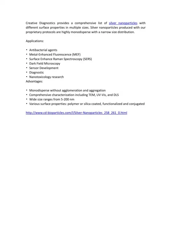
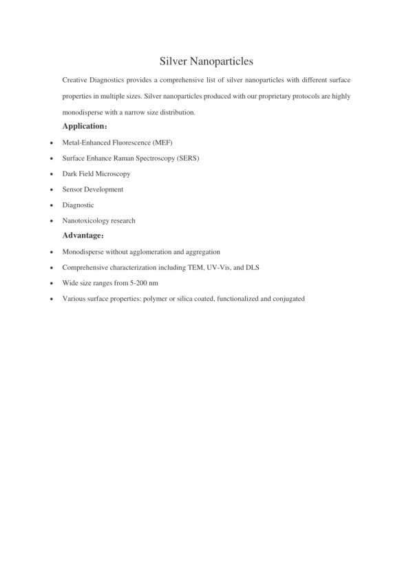
Nanoparticles are being viewed as fundamental building blocks of nanotechnology. The development of new chemical or physical methods, the concern for environmental contaminations are also heightened as the chemical procedures involved in the synthesis of nanoparticles generate a large amount of hazardous byproducts. Thus, there is a need for green chemistry that includes a clean, non toxic and environment friendly method of nanoparticles synthesis. As a result, researchers in the field of nanoparticles synthesis and assembly have turned to biological system of inspiration. One of them is the synthesis of nanoparticles using plant leaf extracts eliminating the elaborate process of marinating the microbial culture and often found to be kinetically favourable than other bioprocesses. The present study deals with the synthesis of silver nanoparticles using leaf extract of Delonix elata. The synthesized silver nanoparticles were predominately spherical in shape, polydispersed and ranged in size from 20 60 nm. K. Vinoth Kumar "Biosynthesis of Silver (Ag) Nanoparticles using Plant Derivatives of Delonix elata" Published in International Journal of Trend in Scientific Research and Development (ijtsrd), ISSN: 2456-6470, Volume-2 | Issue-4 , June 2018, URL: https://www.ijtsrd.com/papers/ijtsrd12910.pdf Paper URL: http://www.ijtsrd.com/other-scientific-research-area/enviormental-science/12910/biosynthesis-of-silver-ag-nanoparticles-using-plant-derivatives-of-delonix-elata/k-vinoth-kumar<br>

E N D
International Research Research and Development (IJTSRD) International Open Access Journal Biosynthesis of Silver (Ag) Nanoparticles sing Plant Derivatives of Delonix elata International Journal of Trend in Scientific Scientific (IJTSRD) International Open Access Journal ISSN No: 2456 ISSN No: 2456 - 6470 | www.ijtsrd.com | Volume 6470 | www.ijtsrd.com | Volume - 2 | Issue – 4 Biosynthes using Plant Derivatives of f Silver (Ag) Nanoparticles K. Vinoth Kumar Department of Environmental Sciences, Department of Environmental Sciences, Tamil Nadu Agricultural University, Tamil Nadu Agricultural University, Coimbatore, Tamil Nadu, India Coimbatore, Tamil Nadu, India ABSTRACT Nanoparticles are being viewed as fundamental building blocks of nanotechnology. The development of new chemical or physical methods, the concern for environmental contaminations are also heightened as the chemical procedures involved in the synt nanoparticles generate a large amount of hazardous byproducts. Thus, there is a need for green chemistry that includes a clean, non toxic and environment friendly method of nanoparticles synthesis. As a result, researchers in the field of nanopart synthesis and assembly have turned to biological system of inspiration. One of them is the synthesis of nanoparticles using plant leaf extracts eliminating the elaborate process of marinating the microbial culture and often found to be kinetically favourable than other bioprocesses. The present study deals with the synthesis of silver nanoparticles using leaf extract of Delonix elata. The synthesized silver nanoparticles were predominately spherical in shape, polydispersed and ranged in size from 20-60 nm. generate a large amount of hazardous byproducts. e is a need for green chemistry that includes a clean, non toxic and environment friendly method of nanoparticles synthesis. As a result, researchers in the field of nanoparticles synthesis and assembly have turned to biological system of inspiration. Among the biological system plants have found application particularly in metal nanoparticles synthesis. Use of plants for synthesis of nanoparticles could be advantageous over other environmentally benign biological processes as this eliminates the elaborate process o maintaining cell culture. Biosynthetic processes for nanoparticles would be more useful if nanoparticles were produced extracelluarly using plants or their extracts and in a controlled manner according to their size, shape and dispersity (Kumar generate a large amount of hazardous byproducts. Thus, there is a need for green chemistry that includes a clean, non toxic and environment friendly method of nanoparticles synthesis. As a result, researchers in the field of nanoparticles synthesis and assembly have turned to biological system of inspiration. Amon biological system plants have found application particularly in metal nanoparticles synthesis. Use of plants for synthesis of nanoparticles could be advantageous over other environmentally benign biological processes as this eliminates the elaborate process o maintaining cell culture. Biosynthetic processes for nanoparticles would be more useful if nanoparticles were produced extracelluarly using plants or their extracts and in a controlled manner according to their size, shape and dispersity (Kumar and Yadav, 2008). Nanoparticles are being viewed as fundamental building blocks of nanotechnology. The development of new chemical or physical methods, the concern for environmental contaminations are also heightened as the chemical procedures involved in the synthesis of nanoparticles generate a large amount of hazardous byproducts. Thus, there is a need for green chemistry that includes a clean, non toxic and environment friendly method of nanoparticles synthesis. As a result, researchers in the field of nanoparticles synthesis and assembly have turned to biological system of inspiration. One of them is the synthesis of nanoparticles using plant leaf extracts eliminating the elaborate process of marinating the microbial culture vourable than other bioprocesses. The present study deals with the synthesis of silver nanoparticles using leaf extract of . The synthesized silver nanoparticles were predominately spherical in shape, polydispersed Biosynthesis of nanoparticles by plant extracts is currently under exploitation. The aqueous silver nitrate solution, after reacting with geranium ) leaf extract, led to rapid formation of highly stable, crystalline silver nanoparticles (16 to 40 nm) (Shankar et al., 2003). Silver nanoparticles were synthesized by treating Capsicum annuum L. leaf extract, the crystalline phase of the nanoparticles changed from polycrystalline to single crystalline and their size increased with increasing reaction time. Five hours reaction time led to spherical and polycrystalline shaped nanoparticles (10 ± 2 nm) (Li et al., 2007). In this paper, we report on the biosynthesis of pure metallic nanoparticles of silver by the reduction of ions with the leaf extract of Delonix Biosynthesis of nanoparticles by plant extracts is currently under exploitation. The aqueous silver nitrate solution, after reacting with geranium (Pelargonium graveolens) leaf extract, led to rapid formation of highly stable, crystalline nanoparticles (16 to 40 nm) (Shankar Silver nanoparticles were synthesized by treating silver ions with Capsicum annuum crystalline phase of the nanoparticles changed from polycrystalline to single crystalline an increased with increasing reaction time. Five hours reaction time led to spherical and polycrystalline shaped nanoparticles (10 ± 2 nm) (Li this paper, we report on the biosynthesis of pure metallic nanoparticles of silver by aqueous Ag+ ions with the leaf extract of elata. Keywords: Green approach, Biosynthesis, Delonix elata, Ag nanoparticles Green approach, Biosynthesis, Delonix INTRODUCTION Nanoparticles are being viewed as fundamental building blocks of nanotechnology. An important aspect of nanotechnology concerns the development of experimental processes for the synthesis of nanoparticles of different sizes, shape and controlled dispersity. With the development of new chemical or physical methods, the concern for environmental contaminations are also heightened as the chemical procedures involved in the synthesis of nanoparticles procedures involved in the synthesis of nanoparticles Nanoparticles are being viewed as fundamental building blocks of nanotechnology. An important aspect of nanotechnology concerns the development of experimental processes for the synthesis of nanoparticles of different sizes, shape and controlled . With the development of new chemical or physical methods, the concern for environmental contaminations are also heightened as the chemical @ IJTSRD | Available Online @ www.ijtsrd.com @ IJTSRD | Available Online @ www.ijtsrd.com | Volume – 2 | Issue – 4 | May-Jun Jun 2018 Page: 83
International Journal of Trend in Scientific Research and Development (IJTSRD) ISSN: 2456-6470 D- Average crystallite size: K- Constant: λ- X-ray Wavelength: β- Angular FWHM of the XRD peak at the diffraction angle: θ- Diffraction angle. MATERIALS AND METHODS Preparation of leaf extract The fresh and young leaf samples of Delonix elata was collected and washed thoroughly with sterile double distilled water (DDW) and finally surface sterilized with 0.1 % HgCl2 for 2 - 3 min under the hood of laminar air flow. Twenty gram of sterilized leaf samples were taken and cut into small pieces. Finely cut leaves were placed in a 500 ml Erlenmeyer flask containing 100 ml of sterile DDW. After that the mixture was boiled for 5 min and filtered. The extract was stored in 4 0C. SEM analysis The thin film of the samples were prepared on a small aluminum plate by just dropping a very small amount of the sample on the plate, extra solution were removed using a blotting paper and then the film on the plate was allowed to dry for overnight. The SEM analysis was performed on a JEOL, model JSM-6390 instrument operated at an accelerating voltage of 20 keV and counting time of 100 s. Synthesis of silver nanoparticles FT-IR measurement Silver nitrate was used as precursor of synthesizing the silver nanoparticles. Five ml of leaf extract was added to 100 ml of 1 mM AgNO3 (99.99 %) aqueous solution in conical flask of 250 ml content at room temperature. The flask was thereafter put into shaker (150 rpm) at 300 C and reaction was carried out for a period of 48 h. FT-IR measurement of sample was performed using the Nicolet Avatar Model FT-IR spectrophotometer in a diffuse reflectance mode at a resolution of 4 cm-1 in KBr pellets. RESULT AND DISCUSSION The extracellular synthesis of silver nanoparticles occurred during the exposure of Delonix elata leaf extract to 1 mM aqueous silver nitrate solution. The complete reduction of silver ions was observed after 48 h of reaction at 300 C under shaking condition. The colour change in reaction mixture was observed during the incubation period, because the formation of silver nanoparticles is able to produce particular colour in the reaction mixtures due to their specific properties. The appearance of dark yellowish-brown colour is a clear indication of the formation of silver nanoparticles in the reaction mixture (fig.1). The colour exhibited by metallic nanoparticles is due to the coherent excitation of all the “free” electrons within the conduction band, leading to an in-phase oscillation and is known as Surface Plasmon Resonance-SPR (Akanna et al., 2010). UV-visible spectroscopy analysis The colour change in reaction mixture was recorded through visual observation. The bioreduction of silver ions in aqueous solution was monitored by periodic sampling of aliquot (1 ml) and subsequently measuring UV-vis spectra of the solution. UV-vis spectra of these aliquot was monitored as a function of time of reaction spectrophotometer (model S3-159) operated at a resolution of 1 nm. on Elico UV-vis XRD measurement The sample was drop-coated onto aluminum plate by just dropping a small amount of sample on the plate frequently, allowed to dry and finally thick coat of sample on plate was prepared. The XRD measurement was performed on a Shimazdu, model LabX-XRD- 6000 instrument operated at a voltage of 20 to 30 keV and a current of 30 mA with Cu K α radiation with a wavelength of 1.5418 Å. UV-vis spectroscopy analysis showed that the SPR absorbance band of silver nanoparticles synthesized using Delonix elata leaf extract centered at 448 nm (fig 2.) and steadily increases in intensity as a function of time of reaction without any shift in the peak wavelength. The frequency and width of the surface plasmon absorption depends on the size and shape of the metal nanoparticles as well as on the dielectric constant of the metal itself and the surrounding medium (Mukherjee et al., 2002). XRD pattern obtained for silver nanoparticles showed characteristic peaks near the 2θ value of 38.760 (fig.3). A Bragg Determination of crystalline size Average crystallite size of silver was calculated using the Scherrer’s formula, D = kλ / βcosθ @ IJTSRD | Available Online @ www.ijtsrd.com | Volume – 2 | Issue – 4 | May-Jun 2018 Page: 84
International Journal of Trend in Scientific Research and Development (IJTSRD) ISSN: 2456-6470 reflection corresponding to the (111) sets of lattice planes are observed which may be indexed based on the face-centered cubic (fcc) structure of silver (Dubey et al., 2009). The XRD pattern thus clearly shows that the silver nanoparticles are crystalline in nature. In addition to the Bragg peak representative of fcc silver nanocrystals, additional and yet unassigned peaks were also observed suggesting that the crystallization of bio-organic phase occurs on the surface of the silver nanoparticles (Sathyavathi et al., 2010). Crystallite size of silver nanoparticles as estimated from the Full width at half maximum (FWHM) of the (111) peak of silver using the Scherrer’s formula exhibited average particles size of 22 nm. Crystalline size of synthesized silver nanoparticles Average Particle size [nm] θ value d- spacing [Å] FWHM [degree] Intensity [CPS] Plant extract [degree] Delonix elata 19.38 2.321 0.703 33.0 21.83 SEM image has shown individual silver particles as well as a number of aggregates. The morphology of the silver nanoparticles was predominately spherical and aggregated into larger irregular structure with no well-defined morphology observed in the micrograph (fig.4). The nanoparticles were not in direct contact even within the aggregates, indicating stabilization of the nanoparticles by a capping agent (proteins secreted by plant leaf extracts). The presence of secondary materials capping nanoparticles and this capping may be assigned to bio-organic compounds from leaf extracts (Rajesh et al., 2009). ethers, carboxylic acids, esters and anhydrides. FT-IR analysis reveals that the carbonyl group from amino acid residues and proteins has the stronger ability to bind metal indicating that the proteins could possibly form a layer covering the metal nanoparticles (i.e., capping of silver nanoparticles) agglomeration and thereby stabilize the medium. This suggests that the biological molecules could possibly perform dual functions of formation and stabilization of silver nanoparticles in the aqueous medium (Sathyavathi et al., 2010). to prevent with the silver CONCLUSION Delonix nanoparticles extracellularly and the synthesized silver nanoparticles are quite stable in solution. The control of shape and size of silver nanoparticles seems to be easy with the use of plant leaf extracts. The synthetic methods based on naturally occurring biomaterials provide an alternative means for obtaining the nanoparticles. Use of plants in synthesis of nanoparticles is quite novel leading to truly ‘green chemistry’ route. This green chemistry approach towards the synthesis of nanoparticles has many advantages such as, ease with which the process can be scaled up, economic viability, eco-friendly and safe way to produce nanoparticles. . elata capable of producing silver Fourier Transform Infra-Red (FT-IR) spectroscopy analysis showed that nanoparticles are capped compounds which are responsible for reduction of silver ions. The wavenumber or frequency (cm-1) of absorption band or peak assigned to the type of vibration, intensity and functional groups of the silver nanoparticles synthesized using Delonix elata leaf extract are shown in fig 5.Different functional groups were involved in reduction of silver ions to silver nanoparticles. The peaks in the region of 3400 to 3200 cm-1 and 3000 to 2850 cm-1 were assigned to O-H stretching of alcohol and phenol compounds and aldehydic -C-H- stretching of alkanes, respectively. The peaks in the region of 1640 to 1550 cm-1 and 1450 to 1375 cm-1 correspond to N-H (bend) of primary and secondary amides and C-H (-CH3 - bend) of alkanes, respectively. The peaks at the region of 1350 to 1000 cm-1 correspond to -C-N- stretching vibration of the amine or -C-O- stretching of alcohols, the synthesized with silver biomolecule REFERENCES 1.Kumar V. and Yadav S.K. (2008). Plant-mediated synthesis of silver and gold nanoparticles and their applications, J. Chem. Technol. Biotechnol., 1, 1- 7. @ IJTSRD | Available Online @ www.ijtsrd.com | Volume – 2 | Issue – 4 | May-Jun 2018 Page: 85
International Journal of Trend in Scientific Research and International Journal of Trend in Scientific Research and Development (IJTSRD) ISSN: 2456 Development (IJTSRD) ISSN: 2456-6470 2.Shankar S.S., Ahmad A., Pasricha R. and Sastry M. (2003). Bioreduction of chloroaurate ions by geranium leaves and its endophytic fungus yields gold nanoparticles of different shapes. Chem., 13, 1822-1826. Shankar S.S., Ahmad A., Pasricha R. and Sastry M. (2003). Bioreduction of chloroaurate ions by geranium leaves and its endophytic fungus yields gold nanoparticles of different shapes. J. Mater. the fungus: Fusarium oxysporum. Chem. Bio. Chem., 3 (5), 461-463. ungus: Fusarium oxysporum. Chem. Bio. 6.Dubey M., Bhadauria S. and Kushwah B.S. (2009). Green synthesis of nanosilver particles from extract of Eucalyptus hybrinda J. Nanomat. Biostruct., 4(3), 537 4(3), 537-543. Dubey M., Bhadauria S. and Kushwah B.S. (2009). Green synthesis of nanosilver particles Eucalyptus hybrinda leaf. Digest 3.Li S., Shen Y., Xie A., Yu X., Qiu L., Zhang L. and Zhang Q. (2007). Green synthesis of silver nanoparticles using Capsicum annuum L. Green Chem., 9, 852-858. Li S., Shen Y., Xie A., Yu X., Qiu L., Zhang L. and Zhang Q. (2007). Green synthesis of silver Capsicum annuum L. extract, 7.Sathyavathi R., Krishna M. B., Rao S.V., Saritha R. and Rao D.N. (2010). Biosynthesis of silver nanoparticles using Coriandrum sativum extract and their application in nonlinear optics. Adv. Sci. Lett., 3, 1-6. rishna M. B., Rao S.V., Saritha R. and Rao D.N. (2010). Biosynthesis of silver Coriandrum sativum leaf extract and their application in nonlinear optics. 4.Akanna S., Prasad K.V., Elumalai E.K. and Savithramma N. (2010). Production of biogen silver nanoparticles using Boswellia ovalifoliolata stem bark, Digest J. Nanomat. Biostruct., 369-372. Akanna S., Prasad K.V., Elumalai E.K. and Savithramma N. (2010). Production of biogenic Boswellia ovalifoliolata 8.Rajesh W.R., Jaya R.L., Niranjan S.K., Vijay D.M. and Sahebrao B.K. (2009). Phytosynthesis of silver nanoparticles using Curr. Nano Sci., 5, 117-122 Rajesh W.R., Jaya R.L., Niranjan S.K., Vijay ahebrao B.K. (2009). Phytosynthesis of silver nanoparticles using Gliricidia sepium. 122 Digest J. Nanomat. Biostruct., 5(2), 5.Mukherjee P., Senapati S., Mandal D., Ahmad A., Khan M.I., Kumar R. and Sastry M. (2002). Extracellular synthesis of gold nanoparticles by Extracellular synthesis of gold nanoparticles by Mukherjee P., Senapati S., Mandal D., Ahmad A., Khan M.I., Kumar R. and Sastry M. (2002). a b c Fig 1. Optical photograph of (a) 1 mM AgNO (b) Leaf extract (c) Leaf (b) Leaf extract (c) Leaf extract + AgNO3 after 48 h of reaction Fig 1. Optical photograph of (a) 1 mM AgNO3 solution 3 peak at 448 nm 2.5 b 2 a Absorbance 1.5 1 0.5 0 330 380 430 480 530 580 630 680 730 Wavelength (nm) a - after 24 h vis spectra of reduction ofFig 3. XRD pattern of Ag nanoparticles Fig 3. XRD pattern of Ag nanoparticles Wavelength (nm) after 24 h Fig 2. UV-vis spectra of reduction of Ag ions to Ag nanoparticles Ag ions to Ag nanoparticles @ IJTSRD | Available Online @ www.ijtsrd.com @ IJTSRD | Available Online @ www.ijtsrd.com | Volume – 2 | Issue – 4 | May-Jun Jun 2018 Page: 86
International Journal of Trend in Scientific Research and International Journal of Trend in Scientific Research and Development (IJTSRD) ISSN: 2456 Development (IJTSRD) ISSN: 2456-6470 Fig 4. SEM image of Ag nanoparticles Ag nanoparticles Fig 4. SEM image of Ag nanoparticles Fig 5. FT-IR spectrum of @ IJTSRD | Available Online @ www.ijtsrd.com @ IJTSRD | Available Online @ www.ijtsrd.com | Volume – 2 | Issue – 4 | May-Jun Jun 2018 Page: 87