Download
1 / 5
50 likes | 59 Views
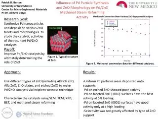
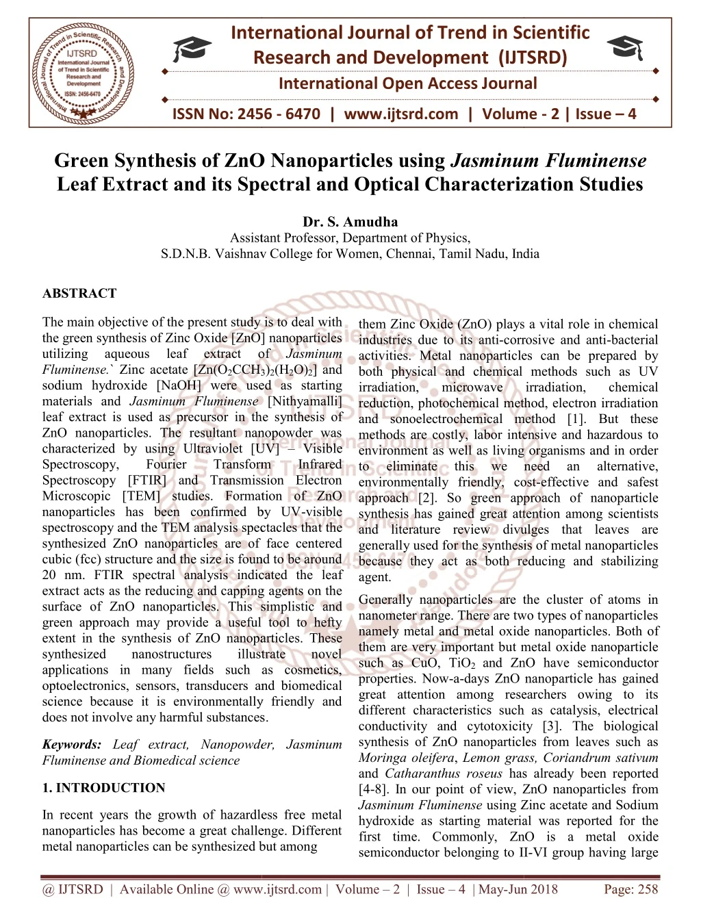
The main objective of the present study is to deal with the green synthesis of Zinc Oxide ZnO nanoparticles utilizing aqueous leaf extract of Jasminum Fluminense.` Zinc acetate Zn O2CCH3 2 H2O 2 and sodium hydroxide NaOH were used as starting materials and Jasminum Fluminense Nithyamalli leaf extract is used as precursor in the synthesis of ZnO nanoparticles. The resultant nanopowder was characterized by using Ultraviolet UV Visible Spectroscopy, Fourier Transform Infrared Spectroscopy FTIR and Transmission Electron Microscopic TEM studies. Formation of ZnO nanoparticles has been confirmed by UV visible spectroscopy and the TEM analysis spectacles that the synthesized ZnO nanoparticles are of face centered cubic fcc structure and the size is found to be around 20 nm. FTIR spectral analysis indicated the leaf extract acts as the reducing and capping agents on the surface of ZnO nanoparticles. This simplistic and green approach may provide a useful tool to hefty extent in the synthesis of ZnO nanoparticles. These synthesized nanostructures illustrate novel applications in many fields such as cosmetics, optoelectronics, sensors, transducers and biomedical science because it is environmentally friendly and does not involve any harmful substances. S. Amudha "Green Synthesis of ZnO Nanoparticles using Jasminum Fluminense Leaf Extract and its Spectral and Optical Characterization Studies" Published in International Journal of Trend in Scientific Research and Development (ijtsrd), ISSN: 2456-6470, Volume-2 | Issue-4 , June 2018, URL: https://www.ijtsrd.com/papers/ijtsrd12908.pdf Paper URL: http://www.ijtsrd.com/physics/nanotechnology/12908/green-synthesis-of-zno-nanoparticles-using-jasminum-fluminense-leaf-extract-and-its-spectral-and-optical-characterization-studies/s-amudha<br>

E N D
International Research Research and Development (IJTSRD) International Open Access Journal International Journal of Trend in Scientific Scientific (IJTSRD) International Open Access Journal ISSN No: 2456 ISSN No: 2456 - 6470 | www.ijtsrd.com | Volume www.ijtsrd.com | Volume - 2 | Issue – 4 Green Synthesis of ZnO Nanoparticles using Leaf Extract and its Spectral and Optical Characterization Studies Leaf Extract and its Spectral and Optical Characterization Studies Leaf Extract and its Spectral and Optical Characterization Studies Green Synthesis of ZnO Nanoparticles using Jasminum Fluminense Jasminum Fluminense Dr. S. Amudha Assistant Professor, Department of Physics, S.D.N.B. Vaishnav College for Women, S.D.N.B. Vaishnav College for Women, Chennai, Tamil Nadu, India Assistant Professor, Department of Physics, Tamil Nadu, India ABSTRACT The main objective of the present study the green synthesis of Zinc Oxide [ZnO] nanoparticles utilizing aqueous leaf Fluminense.` Zinc acetate [Zn(O2CCH3 sodium hydroxide [NaOH] were used as starting materials and Jasminum Fluminense leaf extract is used as precursor in the synthesis of ZnO nanoparticles. The resultant nanopowder was characterized by using Ultraviolet [UV] Spectroscopy, Fourier Spectroscopy [FTIR] and Transmission Electron Microscopic [TEM] studies. Formation of ZnO nanoparticles has been confirmed by UV spectroscopy and the TEM analysis spectacles tha synthesized ZnO nanoparticles are of face centered cubic (fcc) structure and the size is found to be around 20 nm. FTIR spectral analysis indicated the leaf extract acts as the reducing and capping agents on the surface of ZnO nanoparticles. This sim green approach may provide a useful tool to hefty extent in the synthesis of ZnO nanoparticles. synthesized nanostructures applications in many fields such as cosmetics, optoelectronics, sensors, transducers and biomedica science because it is environmentally friendly and does not involve any harmful substances does not involve any harmful substances. present study is to deal with xide [ZnO] nanoparticles leaf them Zinc Oxide (ZnO) plays a vital role in chemical corrosive and anti-bacterial activities. Metal nanoparticles can be prepared by both physical and chemical methods such as UV irradiation, microwave reduction, photochemical method, electron irradiation and sonoelectrochemical method [1]. But these s are costly, labor intensive and hazardous to environment as well as living organisms and in order to eliminate this we environmentally friendly, cost-effective and safest approach [2]. So green approach of nanoparticle gained great attention among scientists and literature review divulges that leaves are generally used for the synthesis of metal nanoparticles because they act as both reducing and stabilizing them Zinc Oxide (ZnO) plays a vital role in chemical industries due to its anti-corrosive and anti activities. Metal nanoparticles can be prepared by both physical and chemical methods such as UV irradiation, microwave reduction, photochemical method, electron irradiation and sonoelectrochemical method [1]. But these methods are costly, labor intensive and hazardous to environment as well as living organisms and in order to eliminate this we environmentally friendly, cost approach [2]. So green approach of nanoparticle synthesis has gained great attention among scientists and literature review divulges that leaves are generally used for the synthesis of metal nanoparticles because they act as both reducing and stabilizing agent. lizing aqueous extract extract of of 3)2(H2O)2] and used as starting [Nithyamalli] in the synthesis of Jasminum irradiation, irradiation, chemical chemical ZnO nanoparticles. The resultant nanopowder was Ultraviolet [UV] – Visible Transform Transform Infrared Infrared need need an an alternative, alternative, IR] and Transmission Electron Formation of ZnO nanoparticles has been confirmed by UV-visible spectroscopy and the TEM analysis spectacles that the icles are of face centered structure and the size is found to be around indicated the leaf as the reducing and capping agents on the Generally nanoparticles are the cluster of atoms in nanometer range. There are two types of nanoparticles namely metal and metal oxide nanoparticle them are very important but metal oxide nanoparticle such as CuO, TiO2 and ZnO have semi properties. Now-a-days ZnO nanoparticle has great attention among researchers owing to its different characteristics such as conductivity and cytotoxicity [3 synthesis of ZnO nanoparticles from leaves such as Moringa oleifera, Lemon grass, C and Catharanthus roseus has already been reported [4-8]. In our point of view, ZnO nanoparticles from Jasminum Fluminense using Zinc acetate and Sodium hydroxide as starting material first time. Commonly, ZnO is a metal oxide semiconductor belonging to II semiconductor belonging to II-VI group having large Generally nanoparticles are the cluster of atoms in nanometer range. There are two types of nanoparticles tal oxide nanoparticles. Both of ut metal oxide nanoparticle This simplistic and green approach may provide a useful tool to hefty synthesis of ZnO nanoparticles. These synthesized nanostructures applications in many fields such as cosmetics, illustrate illustrate novel novel ZnO have semiconductor days ZnO nanoparticle has gained and biomedical great attention among researchers owing to its uch as catalysis, electrical conductivity and cytotoxicity [3]. The biological synthesis of ZnO nanoparticles from leaves such as Lemon grass, Coriandrum sativum has already been reported science because it is environmentally friendly and Keywords:Leaf extract, Nanopowder, Fluminense and Biomedical science Nanopowder, Jasminum 1. INTRODUCTION ZnO nanoparticles from using Zinc acetate and Sodium as starting material was reported for the ZnO is a metal oxide In recent years the growth of hazardless free metal nanoparticles has become a great challenge. Different metal nanoparticles can be synthesized but among metal nanoparticles can be synthesized but among In recent years the growth of hazardless free metal nanoparticles has become a great challenge. Different @ IJTSRD | Available Online @ www.ijtsrd.com @ IJTSRD | Available Online @ www.ijtsrd.com | Volume – 2 | Issue – 4 | May-Jun Jun 2018 Page: 258
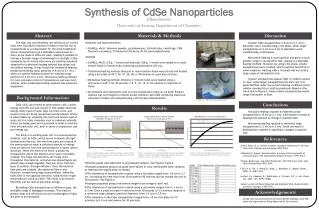
International Journal of Trend in Scientific Research and Development (IJTSRD) ISSN: 2456-6470 stirred at 50 C for another 1 hr until the colloidal solution is obtained. The colour of the resultant solution changes to light pale yellow colour which confirms the presence of ZnO nanoparticles. The precipitate was centrifuged at 10000 rpm at 50 °C for 20 min and powdered specimen was collected. This yellow coloured sample was dried using a hot air oven operating at 70 °C for 2 h and crushed using ceramic mortar and pestle to get fine Zinc Oxide (ZnO) nanoparticles and stored in air-tight bottles for further characterization studies [10]. exciton binding energy of 60 meV and wide band gap of about 3.4 eV. It has wide application in solar cells, gas sensors, ceramics, catalysts and used as an additive in paints, cosmetics, plastics, rubber manufacturing, electronics, agriculture and aquaculture [9]. The plant Jasminum Fluminense belonging to the Olive family called Oleaceae and it contains about 200 species native to tropical and warm temperate regions of Eurasia, Australasia and Oceania. It is a climber plant belonging to dicotyledon and angiosperm group. The main aim of the present study is to synthesize Zinc Oxide (ZnO) nanoparticles using aqueous leaf extract of Jasminum Fluminense and to evaluate the structure by various characterization tools such as Ultraviolet [UV] – Visible Spectroscopy, Fourier Transform Infrared Spectroscopy [FTIR] and Transmission Electron Microscopic [TEM] studies. pharmaceuticals, 2.4 Characterization The optical nanoparticles were investigated using UV-visible 8500 spectrophotometer in the wavelength range 250- 1000 nm. The FTIR spectra were recorded using KBr pellet method by FTIR Spectrophotometer (Bruker, Tensor 27) at the wave number resolution of 1 cm-1 and with a total number of scans as 32 in the range 4000-650 cm-1 on the transmittance mode. The morphology was observed by Transmission Electron Microscopy using Hitachi Model TEM. properties of synthesized ZnO 2.MATERIALS AND METHODS 2.1 Materials Required Zinc acetate [Zn hydroxide [NaOH] pellet and glasswares were purchased from Merck and used as received and Jasminum Fluminense leaves were collected from SDNB Vaishnav College Campus. The leaves and all the glasswares were thoroughly washed with double distilled water before use. 2.2 Preparation of Jasminum Fluminense Leaf Extract Fresh leaves ofJasminum Fluminense about 10 g were collected and washed several times with tap water and then with distilled water and cut into small pieces. These leaves were boiled with 100 ml of double distilled water at 60 C for about 30 minutes. After boiling, color of the solution changes to light brown color and it was cooled at room temperature. This extract was filtered through Whatman Number-1 filter paper and stored in refrigerator for further characterization studies. (O2CCH3)2(H2O)2], Sodium 3.RESULTS AND DISCUSSION 3.1 UV-visible Spectroscopic Results 289.9 nm ZnO nanoparticles 0.5 0.4 Absorbance (A) 0.3 0.2 0.1 0.0 200 300 400 500 600 700 800 900 1000 W avelength (nm ) Fig. 1 UV-visible spectrum of biosynthesized ZnO nanoparticles Figure 1 shows the UV-visible absorption spectrum obtained for the synthesized Zinc Oxide (ZnO) nanoparticles recorded between the range 250-1000 nm. It is known that the ZnO nanoparticles have free electrons due to which Surface Plasmon Resonance (SPR) absorption band appear at 289.9 nm. This absorption peak indicates the reduction of Zn2+ ions in the reaction medium which authenticates the formation of ZnO nanoparticles [4, 11]. No other peaks have been observed which confirms the 2.3 Green Synthesis of ZnO Nanoparticles For the synthesis of ZnO nanoparticles 1mM Zinc acetate [Zn (O2CCH3)2 (H2O) 2] was dissolved in 50 ml of distilled water and kept in stirrer for 1 hr respectively. Then 1mM of Sodium hydroxide [NaOH] Pellets was dissolved in 20 ml of distilled water and kept in stirrer for 1 hr respectively. Then 25 ml of Jasminum Fluminense leaf extract was added drop wise to the above mixture and continuously @ IJTSRD | Available Online @ www.ijtsrd.com | Volume – 2 | Issue – 4 | May-Jun 2018 Page: 259
International Journal of Trend in Scientific Research and Development (IJTSRD) ISSN: 2456-6470 2856 cm-1 are due to C-H stretching vibration [12]. The band at 1750 cm-1 corresponds to C=O stretching [9] whereas the peak at 1561 cm-1 is attributed to asymmetric stretching band from –COO- groups of acetate ions respectively [13]. The intense vibrational band around 1401 cm-1 is assigned to symmetric stretching of carbonyl side group [14] whereas the band at 922 and 858 cm-1 may correspond to O-H bending vibrations of carboxylic acid [15] and C-H bending vibration [10]. The vibrational band observed at 708 and 666 cm-1 corresponds to –C=C-H [16] and M-O stretching of ZnO [17] nanoparticles. The FTIR spectra thus demonstrates the structural changes taking place in the ZnO nanoparticles and Jasminum Fluminense leaf extract by the co-ordination of O-H, C-H, C=O and –COO- bonds. presence of ZnO nanoparticles only. From the UV- visible graph the energy band gap is calculated using the formula The energy band gap value is found to be 4.28 eV which is more than pure ZnO nanoparticles i.e., 3.4 eV. 3.2 FTIR Spectral Analysis of ZnO Nanoparticles FTIR spectral analysis was carried out to find the functional groups present in ZnO nanoparticles and the FTIR spectra of ZnO nanoparticles is shown in Figure 2. The spectrum portraying band at 2924 and Fig. 2 FTIR spectra obtained for biosynthesized ZnO nanoparticles 3.3 TEM Results Fig. 3 TEM image of synthesized ZnO nanoparticles at different magnifications @ IJTSRD | Available Online @ www.ijtsrd.com | Volume – 2 | Issue – 4 | May-Jun 2018 Page: 260
International Journal of Trend in Scientific Research and Development (IJTSRD) ISSN: 2456-6470 The morphology and size of Zinc Oxide (ZnO) nanoparticles examined using Transmission Electron Microscopic (TEM) analysis as shown in Figure 3. These images depicts that the nanoparticles are hexagonal, rod shaped and some of them are spherical at different magnifications. These spherical structures indicate the presence of amorphous nature of ZnO nanoparticles [2]. The average diameter of the ZnO nanoparticle is found to be 20 nm which is correlated with the earlier reports [18]. CONCLUSIONS In this present study eco-friendly, easy synthesis, low- cost, non-hazardous, organically effective and innovative approach of the biosynthesized ZnO nanoparticles using Jasminum Fluminense leaf extract have been reported. The phytochemicals present in the leaf extract acts as a biological stabilizing and reducing agent for the synthesis of metal oxide nanoparticles. The presence of ZnO nanoparticles was confirmed using UV-visible, FTIR and TEM studies. The absorption band observed at 289.9 nm with an energy band gap of 4.28 eV is confirmed by UV- visible spectroscopic studies. FTIR result confirms the presence of functional groups in ZnO nanoparticles. The particle size was found to be around 20 nm with hexagonal Wurtzite structure as evidenced from TEM results. Thus, it is exemplified that the ZnO nanoparticles synthesized in this research work acts as a promising candidate applications. characterization of zno nanoparticles using Moringa Oleifera extract by green synthesis method”, Asian Journal of Phytomedicine and Clinical Research, Vol. 4, (2016), 121 - 132. 5)T.S. Anvekar, V. Rajendra Chari, H. Kadam, “Green synthesis of ZnO nanoparticles, its characterization and application”, Mater. Sci. Res. India, Vol.14, (2017), 153-157. 6)S.S.M. Hassan, W.I.M.E. Azab, H.R. Ali, M.S.M. Mansour, “Green synthesis and characterization of ZnO nanoparticles for photocatalytic degradation of anthracene”, Adv. Nat. Sci.: Nanosci. Nanotechnol., Vol. 6, (2015), 045012. 7)S. Goutam, A.K.Yadav, A. Jyoti Das, “Coriander extract mediated green synthesis of zinc oxide nanoparticles and their structural, optical and antibacterial properties”, J. Nanosci. Tech., Vol. 3, (2017), 249-252. 8)G. Bhumi, N. Savithramma, “Biological synthesis of zinc oxide Catharanthusroseus G. Don. Leaf extract and validation for antibacterial activity”, Int. J. Drug Dev. & Res., Vol. 6, (2014), 208-214. nanoparticles from 9)S. Narendhran, R. Sivaraj, “Biogenic ZnO nanoparticles synthesized using L. aculeata leaf extract and their antifungal activity against plant fungal pathogens”, Bull. Mater. Sci., Vol. 39, (2016), 1–5. for future biological 10)J. Santhoshkumar, Rajeshkumar, nanoparticles using plant leaf extract against urinary tract infection pathogen”, Resource Efficient Technologies, Vol. 3, (2017), 1–7. S.Venkat Kumar, zinc S. “Synthesis of oxide REFERENCES 1)Y.Y. Loo, B.W. Chieng, M. Nishibuchi, S. Radu, “Synthesis of silver nanoparticles by using tea leaf extract from Camellia Sinensis”, Int. J. Nanomed., Vol. 7, (2012), 4263-4267. 11)S. Kavitha, M. Dhamodaran, Rajendra Prasad, M. Ganesan, “Synthesis and characterisation of zinc oxide nanoparticles using terpenoid fractions of Andrographis paniculata leaves”, Int. Nano Lett. Vol. 7, (2017), 141–147. 2)J. Suresh, G. Pradheesh, V. Alexramani, M. Sundrarajan, S.I. Hong, “Green synthesis and characterization of zinc oxide nanoparticle using insulin plant (Costus pictus D. Don) and investigation of its antimicrobial as well as anticancer activities”, Adv. Nat. Sci.: Nanosci. Nanotechnol., Vol. 9, (2018), 015008. 12)A. Geetha, R. Sakthivel, J. Mallika, R. Kannusamy, R. Rajendran, “Green synthesis of antibacterial zinc oxide nanoparticles using biopolymer Azadirachta indica Gum”, Oriental J. Chem., Vol. 32, (2016), 955-963. 3) L.F.A. Anand Raj, E. Jayalakshmy, “Effect of zinc oxide nanoparticle produced by Zingiber Officinale against pathogenic bacteria”, J. Chem. Pharmaceutical Sci., Vol. 8, (2015), Issue 1. 13)D.S. Chauhan , C.S.A. Gopal, D. Kumar, N. Mahato, M.A. Quraishi, M.H. Cho, “Microwave induced green synthesis of nanostructured ZnO as promising antibacterial Nanomed., Vol. 2, (2017), 001-005. 4)C. Thirunavukkarasu, R. Archana, S. Sharmila, B. Janarthanan, J. Chandrasekaran, “Preparation and agent”, Glob. J. @ IJTSRD | Available Online @ www.ijtsrd.com | Volume – 2 | Issue – 4 | May-Jun 2018 Page: 261
International Journal of Trend in Scientific Research and Development (IJTSRD) ISSN: 2456-6470 14)C. Joel, M. Sheik Muhideen Badhusha, “Green synthesis of ZnO nanoparticles using Phyllanthus embilica stem extract and their antibacterial activity”, Der Pharmacia Lettre, Vol. 8, (2016), 218-223. extracts”, Int. J. Chem. Tech. Res., Vol. 10, (2017), 271-275. 17)N. Sundaramurthy, C. Parthiban, “Biosynthesis of copper oxide nanoparticles using Pyrus Pyrifolia leaf extract and evolve the catalytic activity”, Int. Res. J. Eng. Tech., Vol. 2, (2015), 332-338. 15)S.P. Rajendran, K. Sengodan, “Synthesis and characterization of zinc oxide and iron oxide nanoparticles using Sesbania grandiflora leaf extract as reducing agent”, J. Nanosci., (2017), Article ID: 8348507. 18)Shamsuzzaman, A. Mashrai, H. Khanam, R.N. Aljawfi, Biological nanoparticles using C. albicans and studying their catalytic performance in the synthesis of steroidal pyrazolines”, Arabian J. Chem., Vol. 10, (2017), S1530-S1536. synthesis of ZnO 16)S.Vennila, S.S. Jesurani, “Eco-friendly green synthesis and characterization of stable ZnO nanoparticle using small Gooseberry fruits @ IJTSRD | Available Online @ www.ijtsrd.com | Volume – 2 | Issue – 4 | May-Jun 2018 Page: 262