Download
1 / 24
240 likes | 397 Views
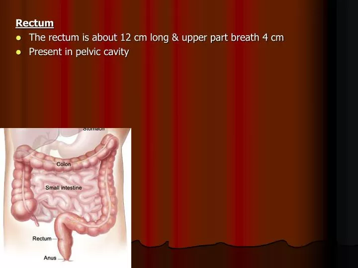
Rectum The rectum is about 12 cm long & upper part breath 4 cm Present in pelvic cavity. Position & Extent begins opposite SV3 as continuation of sigmoid colon passes downwards, following curve of sacrum & coccyx Then extends downwards forward about 2-3 cm in front & below tip of coccyx

E N D
Rectum • The rectum is about 12 cm long & upper part breath 4 cm • Present in pelvic cavity
Position & Extent • begins opposite SV3 as continuation of sigmoid colon • passes downwards, following curve of sacrum & coccyx • Then extends downwards forward about 2-3 cm in front & below tip of coccyx • It abruptly turns downwards & backwards & is continuous with anal canal at anorectal junction
External Apperance The rectum can be distinguished by • absence of mesentery & appendices epiploicae • absence of sacculations • teniae coli to form longitudinal muscle coat
Peritoneum Posterior surface of rectum is entirely non-peritoneal
Posteriorly • Lower 3 pieces of Sacrum, Coccyx & Anococcygeal Ligament • Piriformis, levatorani & coccygeus • Superior, Median & Lower lateral sacral vessels • Sympathetic trunk • Pelvic splanchnic nerves
Interior of Rectum Mucous membrane of empty rectum shows two types of folds Longitudinal fold: - Are transitory. • Present in lower part of empty rectum & obliterated by distension Transverse fold - Permanent • More marked in distended rectum Upper fold – • Near the upper end of rectum & projects from Rt. or Lt. Wall Middle Fold • Largest & most constant lies in upper end of rectal ampulla & projects from anterior & Rt. Walls Lowest Fold • Lies 2.5 cm below middle fold & projects from left wall
Blood Supply Artery • sup rectal art - Continuation of Inferior mesenteric artery • middle rectal art - Branch of Internal Iliac Artery • median sacral art - Branch of Abdominal Aorta
Venous Drainage • follow arteries • however free anastomosis exist between the superior, middle & inferior rectal veins Nerve Supply • Sympathetic from L1, L2 • Parasympathetic from S2-S4
Anal Canal • The anal canal is about 3.8 cm long • begins at level of anorectal junction as a continuation of the rectum • passes downwards & backwards • opens at anal orifice in the perineum Pecularities • Anterior wall of canal is shorter than Posterior wall • Surrounded by Sphincter ani muscles, the tone of which keeps canal closed except during daefecation
Internal Apperance Divided into 3 parts • Upper part = 15 mm • Middle part = 15 mm • Lower part = 8 mm
Upper Part of Anal Canal • lined by mucous membrane (columnar epithelium) • mucosa thrown into vertical folds called anal columns • joined at their lower ends by small semilunar folds called anal valves • The anal valves together form a transverse line that runs all round anal canal called pectinate line
Middle Part • The upper part is separated from the lower part by the pectinate line • Lined by Mucous membrane (stratified squamous epithelium but no sweat & sebaceous gland) • anal columns are absent • Bluishish in Colour due to deep venous plexus • The lower limit of this zone is whitish in appearance so it is referred as white line of Hilton
Lowest part • Cutaneous – lined by skin containing sweat & sebaceous gland
Subcutaneous part • Lies below the level of internal sphincter & surrounds lower part of anal canal • Is in form of flat band about 15 mm broad Superficial part • Surrounds lower part of internal sphincter Deep part • Surrounds upper part of internal sphincter
Blood Supply, Venous Drainage, Lymphatic Drainage & Nerve Supply
Histological Structure of Large Intestine • Mucosa • Mucus surface – Is lined by simple columnar epithelium & numerous goblet cells • Absence of villus • Lamina Propria – Is loaded with numerous deep & straight glands with innumerable mucus goblet cells, lymphatics, smooth muscle fibres & elastic fibres • A thin layer of muscularis mucosa • Sub – mucosa Loose areolar tissue, abundant adipose tissue & blood vessels • Muscularis Inner Circular & outer longitudinal smooth muscle • Serosa - Peritoneum