Download
1 / 25
250 likes | 292 Views
Digestion. Copy everything in red. The Digestive System’s Function. As food passes through the digestive system, it gets broken down distributing its nutrient value to the body

E N D
Digestion Copy everything in red
The Digestive System’s Function • As food passes through the digestive system, it gets broken down distributing its nutrient value to the body • The function of each organ of the digestive system is to help convert foods into simpler molecules that can be absorbed and used by the cells of the body
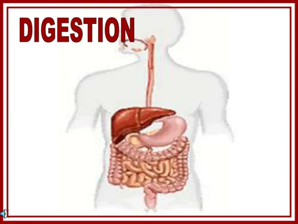
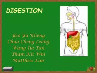
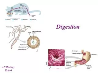
The Digestive System Structures • The digestive system includes: • Mouth • Pharynx • Esophagus • Stomach • Small intestine • Large intestine • Major accessory structures that add secretionsto the digestive system include: • Salivary glands • Pancreas • Liver
Mouth Pharynx Salivary glands Esophagus Liver Stomach Pancreas (behind stomach) Gallbladder (behind liver) Large intestine Small intestine Rectum Figure 38–10 The Digestive System
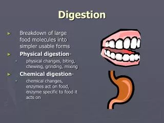
Mouth & Salivary Glands • Teeth • Protected by enamel • Chewing begins the process of mechanical digestion = physical breakdown of large pieces of food into smaller pieces (cutting, tearing, crushing) • Saliva • Secreted by salivary glands • Moisten food making it easier to chew • Begins the process of chemical digestion = Amylase (digestive enzyme) chemically breakdown large starch molecules into smaller sugar molecules • Lysozyme enzyme fights infection
Pharynx • Remember from the Respiratory System: • The pharynx (throat) is a tube in the back of the mouth that passes both air and food • Epiglottis (flap of tissue) covers the trachea to ensure food continue to move along digestive tract
Esophagus • Food tube • Bolus (chewed clump of food) moves along by contractions of smooth muscle surrounding the esophagus = peristalsis • Cardiac sphincter closes esophagus after food has passed into the stomach preventing stomach contents from moving back into the esophagus (heart burn occurs when stomach acid splashing into the esophagus)
Stomach • A large muscular sac made of smooth muscle that mechanically and chemically digests food • Chemical digestion occurs as gastric glands in the stomach lining secrete mucus to protect the inner wall while pepsin and hydrochloric acid break down protein • Ulcers = a hole in the stomach wall caused by a bacterial infection that eats away at the lining of the stomach • Mechanical digestions occurs as the stomach muscles contract to churn and mix stomach fluids producing a mixture = chyme • Pyloric valve opens allowing chyme to flow from the stomach into the small intestine
Duodenum • More chemical digestion of chyme occurs in the duodenum (the first part of the small intestines) • Chyme mixes with enzymes and digestive fluids from the pancreas and the liver (accessory structures)
Pancreas • A gland that produces hormones that regulate blood sugar levels • Produces enzymes that break down carbohydrates, proteins, lipids, and nucleic acids • Produces sodium bicarbonates (a base) that neutralizes stomach acid
Liver • Produces bile = “detergent” dissolving fat droplets • Bile is stored in the gallbladder
Liver Bile duct Pancreas Gallbladder Pancreatic duct Duodenum To small intestine Figure 38–13 The Liver and the Pancreas Section 38-2
Small Intestine • Made of three parts: duodenum, jejunum, and ileum • Where chemical digestion is completed • The folded surfaces are covered with villi = small fingerlike projections that increase the surface area of the small intestines for greater absorption of nutrients • The products of carbohydrate or protein digestion are absorbed into capillaries in the villi and microvilli • Undigested fats are absorbed by lymph vessels = lacteals
Circular folds Epithelial cells Villi Capillaries Lacteal Vein Artery Figure 38–14 The Small Intestine Section 38-2 Villus Small Intestine
The Digestive EnzymesKNOW THIS CHART Section 38-2 Site Mouth Stomach Small intestine(from pancreas) Small intestine Enzyme Role in Digestion Breaks down starches into disaccharides Breaks down proteins into large peptides Continues the breakdown of starch Continues the breakdown of protein Breaks down fat Breaks down remaining disaccharides into monosaccharides Breaks down dipeptides into amino acids. Salivary amylase Pepsin Amylase Trypsin Lipase Maltase, sucrase, lactase Peptidase
Large Intestine • Food entering large intestine is basically nutrient-free, mainly made of water, cellulose (fiber), and other indigestible substances • Removes water left in the chyme by absorbing it across the large intestine wall • If water removal is inefficient, diarrhea occurs and can be dangerous due to the loss of salts and water • Intestinal bacteria help with digestion • The appendix is believed to have formerly stored bacteria to assist with cellulose digestion • Solid concentrated waste (feces) is excreted through the rectum
Excretion Copy everything in red
Excretion • Excretion = the process by which wastes are eliminated from the body • The excretory system includes: • Lungs: excrete gaseous carbon dioxide from cellular respiration • Rectum: excrete solid undigested remainsfrom food • Skin: excretes excess water, salts, urea • Kidneys and accessory organs
The Urinary System • The urinary system rids the blood of wastes produced by the metabolism of nutrients and controls blood volume by removing excess water produced by body cells. • The urinary system includes: • Kidneys • Urinary bladder • Connecting tubules: • Ureter • Urethra
The Urinary SystemDRAW & LABEL THIS Section 38-3 Artery Vein Kidney (Cross Section) Kidney Cortex Medulla Ureter Urinary bladder Urethra
Kidneys • Most people have 2 kidneys located on either side of the spinal column on your lower back • Ureters = tubes that carry urine from each kidney to the urinary bladder • Urinary bladder = saclike organ that stores urine until it can be excreted • The kidneys filter blood by removing urea, excess water and other wastes collected as urine and the clean filtered blood returns to circulation
Kidney Structure • Inner part = renal medulla • Outer part = renal cortex • Functional units of the kidney = nephrons • About 1 million nephrons in each kidney • Each nephron has its own arteriole (small artery), venule (small vein), and network of capillaries to filter blood
Capillaries Bowman’s capsule Cortex Glomerulus Renal artery Medulla Renal vein Collecting duct Ureter Vein To the bladder Artery To the ureter Loop of Henle Figure 38–17 Structure of the Kidneys Section 38-3 Kidney Nephron FILTRATION…REABSORBTION…SECRETION
Urine • The material that remains = urine containing urea, salts, water and other substances • The loop of Henle conserves water and minimizes the volume of urine • Urine is stored in the urinary bladder until it can be released from the body through a tube = urethra
Kidney Function • The kidneys maintain homeostasis by: • Regulating the water content of the blood (blood volume) • Maintaining blood pH • Removing waste products from the blood