Download
1 / 19
190 likes | 197 Views
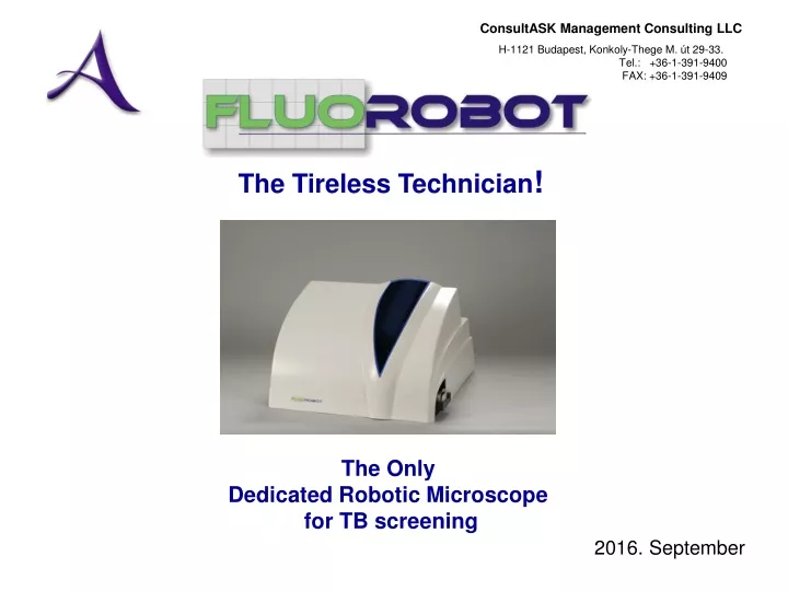
The Tireless Technician !. The Only Dedicated Robotic Microscope for TB screening. 2016. September. The Road behind our development team. Core team has more than 15 years experience in medical imaging and robotic microscopy

E N D
The Tireless Technician! The Only Dedicated Robotic Microscope for TB screening 2016. September
The Road behind our development team • Core team has more than 15 years experience in medical imaging and robotic microscopy • Main project in the early 90s was the Seditron urine sediment analyzer developed and produced for Boehringer Mannheim
First stage of Development1996-1997 • ASK was approached by the Semmelweis University of Medicine Department of Respiratory Medicine in 1995 ( Dr. Ákos Somoskövi, Prof. Magyar) to develop an automated robot microscope with image processing for detection of AFB in fluorescent stained sputum smears • A prototype was developed in 1996, and a clinical study at the University Laboratory was conducted with 132 + 107 smears, parallelly tested with Ziehl-Neelsen method • The results were convincing and were presented at several meetings and published in Int. J. Tuberc Lung Dis, 1999 by the team Conclusions -Technical feasibility was proven -Cost of equipment was relatively high, due to complexity and precision of mechanics (not necessarily needed), traditional fluorescent light source (lifetime) and availability of low cost/high speed electronic components at the time -TB was not in the forefront of interest
The main objectives at relaunch (2009) Develop an instrument that is: • Reliable, robust • Affordable • Small footprint • Has a high walk-away window • Easy-to-use, „foolproof” User Interface • Can be used without change in workflow Eliminate the human factors of reading - tiring effect - different level of expertise - reproducibility, standardisation (path, area?) Enable archiving, revisiting, transmitting of images for: - QC - telediagnosis - elimination of slide storage Open new market segment for follow-up projects -automatic sample preparation
Traps we wish to avoid We are not suggesting new, unknown methodology to be proven • (Smear microscopy with its known advantages/diasadvantages) We do not promise to improve the inherent sensitivity of smear microscopy - (But we can introduce standardisation for example, lacking today) We do not compete with other market segments - X-ray, culture, PCR, … We compete with the human microscopist! We do not want to compete on bacteria level image processing theories, but provide reliable results at sample level We do not sacrifice specificity for virtual increase in sensitivity - if positives have to be re-checked, we do not see economic rationale
Scientfic advisors helping the Fluorobot project • Dr. Ákos SOMOSKÖVI, Scientific Advisor,earned his MD degree in 1993 and his PhD in 1999 from the Albert Szent-Györgyi University of Medical Sciences in Szeged, Hungary. In 2004, he was awarded a Doctor of Science degree by the Hungarian Academy of Sciences. After graduation, he joined the Department of Respiratory Medicine, School of Medicine, Semmelweiss University in Budapest, Hungary where he was appointed Associate Professor in the Department of Respiratory Medicine, School of Medicine. Between 2001 and 2002, Dr.Somoskövi was a research fellow at the Bloomberg School of Public Health, Johns Hopkins University, Baltimore, Maryland. Between 2005 and 2006, he also had the privilege to be Associate Director of the Clinical Mycobacteriology Laboratory, Wadsworth Center, New York State Department of Health in Albany, N.Y. Until joining the Foundation for Innovative New Diagnostics (FIND, Geneva) in 2009, he worked in the International Laboratory Branch of the Global AIDS Program at the CDC in Atlanta. After he was working for the Swiss National Reference Center for Mycobacteria in Zurich.an today he is senior consultant physician at the Department of Respiratory Medicine of Skaraborg Hospital (Skövde, Sweden). Dr. Somoskövi is Board Certified in respiratory medicine and clinical oncology, with broad experience in clinical management, epidemiology and laboratory diagnosis of tuberculosis. Dr Sabine Rüsch-Gerdes has been working in the department of Mycobacteria at the Forschungszentrum Borstel since 1975. She was the head of the National Reference Laboratory for Mycobacteria in Germany and the head of one of the Supranational Reference Laboratories of WHO. She performs External Quality Control for all TB laboratories in Germany and, in addition, for approximately 40 other countries worldwide. For many years, Dr Rüsch-Gerdes has worked as a consultant for different organizations, such as WHO, KNCV, MSF, and ICRC in different countries in Europe (e.g. Kazakhstan, Republic of Moldova, Armenia, Azerbaijan) and Africa (e.g. Uganda, Sierra Leone, Ghana). Nearly all TB diagnostics techniques have been evaluated in the laboratory of Dr Rüsch-Gerdes.
Scientific advisors, FINDFIND Geneva, FIND India Head of TB Programme India & SE Asia Dr. C.N. Paramasivan In April 2006, Dr. Paramasivan (“Param”) joined FIND from his positionasHeadof the Bacteriology Division of the Tuberculosis Research Centre of the Indian Council of Medical Research (ICMR) in Chennai, India. He earned his Ph.D. in Medical Microbiology, as well as a D.Sc. from Madras University. He is also fellow of National academy of Medical Sciences (FAMS) and fellow of National academy of Sciences (FNASc). Param has made several important contributions to Tuberculosis research at the Tuberculosis Research Centre, and is responsible for establishing state-of-the-art laboratory facilities for tuberculosis, acute respiratory illness, opportunistic infections for patients suffering from HIV/TB, and for the HPLC method for species identification of mycobacteria. His contributions also included detailed identification of environmental mycobacteria and their role in the protective response offered by BCG; in vitro and in vivo simulation studies with newer anti tubercular drugs and regimens; and simplifying and standardizing quality assured laboratory diagnosis of tuberculosis. He took active interest in training laboratory personnel, establishing and streamlining TB laboratory services in SEAR and SAARC countries. He also played a major role in the conduction of drug resistance surveillance studies in India and other SEAR countries. From 1990 to 2004, Param served as Head of the WHO Supra National Reference Laboratory for SEAR, and in 2001, the WHO nominated him as Member of the WHO Expert Advisory Panel on Tuberculosis. In 2004, the IUATLD elected him to the Chair of the Bacteriology and Immunology section for their Global Meetings and later as Secretary of TB section and he currently serves as its Programme Secretary. Chief Scientific Officer Dr. Mark Perkins Dr. Mark Perkins trained in Internal Medicine and Pediatrics at Vanderbilt in Nashville, Tennessee, and was chief resident in Pediatrics. He had a research fellowship in the laboratory of Infectious Diseases at the National Institutes of Health in Bethesda, Maryland, where his research focused broadly on the development of respiratory virus vaccines, and specifically on identifying genotypic changes responsible for adaptation to low-temperature growth (influenza) and monoclonal escape (RSV). After returning to Vanderbilt to complete a clinical fellowship in Infectious Diseases, he went to Duke University, first as a Senior Fellow in Clinical Microbiology, and then as a faculty member in the Division of Infectious Diseases and International Health. He worked in Brazil for 5 years to develop and co-direct a collaborative Duke facility for research in tropical diseases and to establish a reference diagnostic laboratory. Dr. Perkins is Board Certified in Internal Medicine, Pediatrics, Infectious Diseases, Clinical Microbiology, and Tropical Medicine. In 1998 he joined the Global Tuberculosis Programme of the World Health Organization and the following year established a Diagnostics unit in the Special Programme for Research and Training in Infectious Diseases (TDR). His group focused on the development and evaluation of new diagnostics for tuberculosis, malaria, schistosomiasis, leishmania, trypanosomiasis and sexually transmitted diseases.
Appearences of Fluorobot following the non-public tests • 2012 MEDICA, Dusseldorf (Concept and video only) • 2013 March, Medical Fair India, New Delhi (early prototype I., booth, demonstration) • 2014 44th Union Meeting on Lung Health, Paris (pre-prototype, booth, demonstration) • 2014 Submission to EU H2020 SME Instrument for funding clinical evaluation
EU SME Instrument EvaluationSubmitted by our technology partner ASK-M
Sample slide from Claudia Denkkinger’s (Head of TB programme, FIND) Presentation on TB diagnostic pipeline (September 2015)
Main features of technology applied • Illumination system using LED technology, less fragile, longer lifetime and cheaper then gas lamp excitation • Cost effective, dedicated optical path • Simplified, more cost effective X-Y movement • Focusing based on mechanic solution (proprietary) eliminating real-time focusing need • Minimized footprint and heat emission , to resist harsh environmental conditions • Standardised computer equipment • Not targeting a conventional, multipurpose, real laboratory microscope design, focusing instead on the most affordable single task solution (one single magnification)
Samples used • Standard Auramine O stained smears • Samples are from the Hungarian TBC Reference Laboratory at Korányi Institute headed by Dr. Nóra Szabó, and FIND/WHO test panels used for calibration, testing and training manual fluorescence microscopy, prepared in Reference Laboratory in India, and the US. • Prototype of dedicated slide with fluorescent focusing mark is used (India)
Main software components • Embedded firmware for hardware related functions and the communication with the main software framework (motors, LED source control, transducers, timings, etc.) • Image processing data input and firmware control software (part of the main framework), such as camera control, integration time, path definition, etc. • Image processing software • AI (artifical Intelligence), i.e. pattern recongition algorithms • UI software for communication, displaying results, file handling, communication, etc. • Maintenance functions for adjustment, debugging, etc.
Main modules of the image processing software • Grabbing and digitising viewfield • Filtering, normalisation of images • Extraction of fields of interest • Localising potential objects • Determining morphological parameters of objects • Decision making of objects in differnet classes
Join the Journey! www.fluorobot.com
Let’s Improve Treatment Chances Further! Aix-Les-Bains, FR