Download
1 / 69
690 likes | 940 Views
Epithelial Tissue. Chapter 4 Anatomy and Physiology Liberty High School Mr. Knowles. Tissue. Collections of specialized cells and cell products that perform a specific function. Four Types of Tissue. Epithelial- covers exposed surfaces, lines passageways, forms glands.

E N D
Epithelial Tissue Chapter 4 Anatomy and Physiology Liberty High School Mr. Knowles
Tissue • Collections of specialized cells and cell products that perform a specific function.
Four Types of Tissue • Epithelial- covers exposed surfaces, lines passageways, forms glands. • Connective- fills internal space, structural support, storage of energy. • Muscle -contracts for specific movements. • Neural- carries information from one part of the body to another.
Observe my Lunch on the Front Table! • List three functions of the plastic bag.
Epithelial Tissue • Includes epithelia and glands. • Epithelium- a layer of cells that forms a barrier. Epithelia- plural, many types; Epithelial is the adjective. • Examples: surface of skin; lining of the digestive, respiratory, reproductive tracts.
Functions of Epithelia • Provide Physical Protection- protect surfaces from abrasion, dehydration, chemical and biological agents. • Control Permeability- regulates molecules that enter or leave through the surface.
Permeability Can Change! Corn Callus
Functions of Epithelia • Provides Sensation- many sensory nerves; Ex: smell, taste, hearing. • Produce Specialized Secretions- gland cells produce secretions
Some Characteristics • Cellularity: tightly bound cells with little space between. • Polarity: has an exposed surface- apical surface (faces exterior surface ) and an attached surface-basal surface (attached to underlying tissue). Organelles are distributed unevenly in these cell.
Characteristics • Attachment: basal surface of an epithelium is bound to a thin basement membrane- produced by the basal surface of epithelium and underlying connective tissue. • Avascularity: epithelia have no blood vessel; epithelial cells receive nutrients by diffusion through apical and basal surfaces.
Characteristics • Regeneration: cells damaged or lost at the apical surface are replaced constantly.
How do epithelial cells do ALL of this? The answer is in their structure! Function Structure
Specialized Epithelial Cells Some epithelial cells: • Produce secretions. • Help with movement of fluids over epithelial surface. • Help move fluids through the epithelium. These cells have a strong polarity (top and bottom).
Apical Surface Structures • Microvillus (i)- small projections of the cell; a few to many on each cell. Function: increase surface area (20X) of epithelial cell (transport specialists). Location: epithelial surfaces where there is absorption and secretion; along digestive and urinary tracts See Fig. 4-1, p. 110.
Apical Surface Structures 2. Cilia- different internal structure than microvilli; many, long extensions that beat in a coordinated fashion. Function: movement of material along the epithelium. Location: respiratory tract (mucus); fallopian tubes (egg)
Apical Surface Structures 3. Stereocilia- similar to microvilli but longer, but cannot move. Function: detection of vibration. Location: male reproductive tract; receptor cells of inner ear.
Stereocilia Stereocilia
Staying Together! 3 Ways Epithelial Cells Stay Together: 1. Intercellular Connections 2. Attachment to Basal Membrane 3. Epithelial Maintenance and Repair
1. Intercellular Connections or Cell Junctions • Tight Junctions- fusion of cell membranes of neighboring cells. Function: block water and solutes between cells; protection. Location: near apical surfaces of cells in digestive tract-keep enzymes and acids from damaging cells underneath.
1. Intercellular Connections or Cell Junctions b. Desmosomes- strong connections of proteins (CAMs) between cells. Function: Act as cross-braces between cells (tent ropes) to hold the shape of cells; anchor cell to it’s base. Found: basal and lateral; superficial skin and cardiac muscle cells; Ex. Dead skin comes off as sheet.
1. Intercellular Connections or Cell Junctions c. Gap Junctions- interlocking membrane proteins (connexons) that form channels between cells. Function: allow small molecules and cations to pass between cells-coordinate functions-beating cilia. Found: lateral; cardiac and smooth muscle
2. Basement Membrane • A layer of protein fibers to which epithelia can attach. • Separates epithelial layer from connective tissue.
3. Maintenance and Repair • Epithelial cells must be replaced due to damage-bacteria, enzymes, toxic chemicals, etc. • Use germinative cells (a type of stem cell) to replace them. • Located in deepest layer near basement membrane.
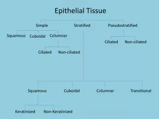
How many kinds of epithelia are there? Classified in Two Ways: Number of Layers and Shape of Cell
A. Layers • Simple- a single layer covers the B.M. Characteristics: thin layer, fragile; no mechanical protection. Location: lines internal passageways and compartments. Ex. Walls of blood vessels; internal surface of lungs. Function: absorption and secretion; reduce friction in vessels.
A. Layers • Stratified- several layers of cells cover the B.M. Characteristics: only one layer contacts the B.M.; other layers lay on top of these cells. Location: surface of the skin; lining of the mouth. Function: protect from mechanical and chemical stress.
B. Shape • Squamous- “scale” thin, flat cells; look like fried eggs laid side by side. Ex. Simple Squamous Epithelium- walls of alveoli of lung; walls of blood vessels and inner heart chamber- called Endothelium. Ex. Stratified Squamous Epithelium- surface of skin; lines mouth, etc.; provides protection.
B. Shape • Cuboidal- hexagonal boxes; nuclei near center of cells. Ex. Simple Cuboidal- mostly secretion and absorption; kidney tubules; salivary glands. Stratified Cuboidal- rare in body; lines the ducts of sweat and mammary glands. Transitional Epithelium- are a type of stratified cuboidal; allows expansion and contraction; lines urinary bladder.