Download
1 / 24
270 likes | 753 Views
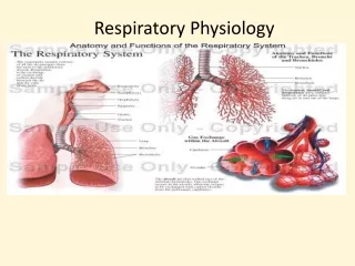
Chapter 24 Physiology of the Respiratory System. Respiratory Physiology. Respiratory physiology—complex, coordinated processes that help maintain homeostasis Respiratory function includes the following: External respiration Pulmonary ventilation (breathing) Pulmonary gas exchange

E N D
Respiratory Physiology • Respiratory physiology—complex, coordinated processes that help maintain homeostasis • Respiratory function includes the following: • External respiration • Pulmonary ventilation (breathing) • Pulmonary gas exchange • Transport of gases by the blood • Internal respiration • Systemic tissue gas exchange • Cellular respiration • Regulation of respiration
Pulmonary Ventilation • Respiratory cycle (ventilation; breathing) • Inspiration—moves air into the lungs • Expiration—moves air out of the lungs • Mechanism of pulmonary ventilation • Pulmonary ventilation mechanism must establish two gas pressure gradients : • One in which the pressure within alveoli of lungs is lower than atmospheric pressure to produce inspiration • One in which the pressure in alveoli of lungs is higher than atmospheric pressure to produce expiration • Pressure gradients are established by changes in size of thoracic cavity that are produced by contraction and relaxation of muscles • Boyle’s law—the volume of gas varies inversely with pressure at a constant temperature
Pulmonary Ventilation • Mechanism of pulmonary ventilation (cont.) • Inspiration—contraction of diaphragm produces inspiration—as it contracts, it makes thoracic cavity larger • Expansion of thorax results in decreased intrapleural pressure (Pip), leading to a decreased alveolar pressure (Palv) • Air moves into lungs when alveolar pressure (Palv) drops below atmospheric pressure (Patm) • Compliance—ability of pulmonary tissues to stretch, making inspiration possible
Pulmonary Ventilation • Mechanism of pulmonary ventilation (cont.) • Expiration—a passive process that begins when inspiratory muscles are relaxed, decreasing size of thorax • Increasing thoracic volume increases intrapleural pressure and thus increases alveolar pressure above atmospheric pressure • Air moves out of lungs when alveolar pressure exceeds atmospheric pressure • Pressure between parietal and visceral pleura is always less than alveolar pressure and less than atmospheric pressure; the difference between Pip and Palv is called transpulmonary pressure • Elastic recoil—tendency of pulmonary tissues to return to a smaller size after having been stretched passively during expiration
Pulmonary Ventilation • Pulmonary volumes—the amounts of air moved in and out and remaining are important to the normal exchange of oxygen and carbon dioxide • Spirometer—instrument used to measure volume of air • Tidal volume (TV)—amount of air exhaled after normal inspiration • Expiratory reserve volume (ERV)—largest volume of additional air that can be forcibly exhaled (between 1.0 and 1.2 liters is normal ERV) • Inspiratory reserve volume (IRV)—amount of air that can be forcibly inhaled after normal inspiration (normal IRV is 3.3 liters) • Residual volume (RV)—amount of air that cannot be forcibly exhaled (1.2 liters)
Pulmonary Ventilation • Pulmonary capacities—the sum of two or more pulmonary volumes • Vital capacity—the sum of IRV + TV + ERV • Minimal volume—amount of air remaining after RV • A person’s vital capacity depends on many factors, including the size of the thoracic cavity and posture • Functional residual capacity—amount of air at the end of a normal respiration • Total lung capacity—the sum of all four lung volumes—the total amount of air a lung can hold
Pulmonary Ventilation • Pulmonary capacities (cont.) • Alveolar ventilation—volume of inspired air that reaches the alveoli • Anatomical dead space—air in passageways that do not participate in gas exchange • Physiological dead space—anatomical dead space plus the volume of any nonfunctioning alveoli (as in pulmonary disease) • Alveoli must be properly ventilated for adequate gas exchange
Pulmonary Ventilation • Pulmonary air flow—rates of air flow into/out of the pulmonary airways • Total minute volume—volume moved per minute (ml/min) • Forced expiratory volume (FEV) or forced vital capacity (FVC)—volume of air expired per second during forced expiration (as a percent of VC) (Figure 24-12) • Flow-volume loop—graph that shows flow (vertically) and volume (horizontally), with top of loop representing expiratory flow-volume and bottom of loop representing inspiratory flow-volume
Pulmonary Gas Exchange • Partial pressure of gases—pressure exerted by a gas in a mixture of gases or a liquid • Law of partial pressures (Dalton’s law)—the partial pressure of a gas in a mixture of gases is directly related to the concentration of that gas in the mixture and to the total pressure of the mixture • Arterial blood Po2 and Pco2 equal alveolar Po2 and Pco2
Pulmonary Gas Exchange • Exchange of gases in the lungs takes place between alveolar air and blood flowing through lung capillaries • Four factors determine the amount of oxygen that diffuses into blood: • The oxygen pressure gradient between alveolar air and blood • The total functional surface area of the respiratory membrane • The respiratory minute volume • Alveolar ventilation
Pulmonary Gas Exchange • Exchange of gases in the lungs (cont.) • Structural factors that facilitate oxygen diffusion from alveolar air to blood: • Walls of the alveoli and capillaries form only a very thin barrier for gases to cross • Alveolar and capillary surfaces are large • Blood is distributed through the capillaries in a thin layer so each red blood cell comes close to alveolar air
How Blood Transports Gases • Oxygen and carbon dioxide are transported as solutes and as parts of molecules of certain chemical compounds • Transport of oxygen • Hemoglobin is made up of four polypeptide chains (two alpha chains, two beta chains), each with an iron-containing heme group; carbon dioxide can bind to amino acids in the chains, and oxygen can bind to iron in the heme groups • Oxygenated blood contains about 0.3 ml of dissolved O2 per 100 ml of blood • Hemoglobin increases the oxygen-carrying capacity of blood • Oxygen travels in two forms: as dissolved O2 in plasma and associated with hemoglobin (oxyhemoglobin) • Increasing blood Po2 accelerates hemoglobin association with oxygen • Oxyhemoglobin carries the majority of the total oxygen transported by blood
How Blood Transports Gases • Transport of carbon dioxide (CO2) • A small amount of CO2 dissolves in plasma and is transported as a solute (10%) • Less than one fourth of blood CO2 combines with NH2 (amine) groups of hemoglobin and other proteins to form carbaminohemoglobin (20%) • Carbon dioxide association with hemoglobin is accelerated by an increase in blood Pco2 • More than two thirds of the carbon dioxide is carried in plasma as bicarbonate ions (70%)
Systemic Gas Exchange • Exchange of gases in tissues takes place between arterial blood flowing through tissue capillaries and cells (Figure 24-27) • Oxygen diffuses out of arterial blood because the oxygen pressure gradient favors its outward diffusion • As dissolved oxygen diffuses out of arterial blood, blood Po2 decreases, which accelerates oxyhemoglobin dissociation to release more oxygen to plasma for diffusion to cells (Figure 24-28)
Systemic Gas Exchange • Carbon dioxide exchange between tissues and blood takes place in the opposite direction from oxygen exchange • Bohr effect—increased Pco2 decreases the affinity between oxygen and hemoglobin (Figure 24-29, A) • Haldane effect—increased carbon dioxide loading caused by a decrease in Po2 (Figure 24-29, B)
Regulation of Pulmonary Function • Respiratory control centers—the main integrators that control the nerves that affect inspiratory and expiratory muscles are located in the brainstem (Figure 24-30) • Medullary rhythmicity center—generates the basic rhythm of respiratory cycle • This area consists of two interconnected control centers: • Inspiratory center stimulates inspiration • Expiratory center stimulates expiration • Basic breathing rhythm can be altered by different inputs to medullary rhythmicity center (Figure 24-30) • Input from apneustic center in pons stimulates inspiratory center to increase length and depth of inspiration • Pneumotaxic center—in pons—inhibits apneustic center and inspiratory center to prevent overinflation of lungs
Regulation of Pulmonary Function • Factors that influence breathing—sensors from the nervous system provide feedback to medullary rhythmicity center (Figure 24-31) • Changes in the Po2, Pco2 and pH of arterial blood influence medullary rhythmicity area • Pco2 acts on central chemoreceptors in medulla—if it increases, result is faster breathing; if it decreases, result is slower breathing • A decrease in blood pH stimulates peripheral chemoreceptors in the carotid and aortic bodies, and even more so, the central chemoreceptors (because they are surrounded by unbuffered fluid) • Arterial blood Po2 presumably has little influence if it stays above a certain level • Arterial blood pressure controls breathing through respiratory pressoreflex mechanism • Hering-Breuer reflexes help control respirations by regulating depth of respirations and volume of tidal air • Cerebral cortex influences breathing by increasing or decreasing rate and strength of respirations
Regulation of Pulmonary Function • Ventilation and perfusion (Figure 24-32) • Alveolar ventilation—air flow to the alveoli • Alveolar perfusion—blood flow to the alveoli • Efficiency of gas exchange can be maintained by limited ability to match perfusion to ventilation—for example, vasoconstricting arterioles that supply poorly ventilated alveoli and allow full blood flow to well-ventilated alveoli
The Big Picture: Respiratory System and the Whole Body • The internal system must continually get new oxygen and rid itself of carbon dioxide because each cell requires oxygen and produces carbon dioxide as a result of energy conversion • Specific mechanisms involved in respiratory function: • Blood gases need blood and the cardiovascular system to be transported between gas exchange tissues of lungs and various systemic tissues of body • Regulation by the nervous system adjusts ventilation to compensate for changes in oxygen or carbon dioxide levels in internal environment • Skeletal muscles of the thorax aid airways in maintaining flow of fresh air • Skeleton houses the lungs, and the arrangement of bones facilitates the expansion and recoil of the thorax • Immune system prevents pathogens from colonizing the respiratory tract and causing infection