Download
1 / 24
350 likes | 1.12k Views
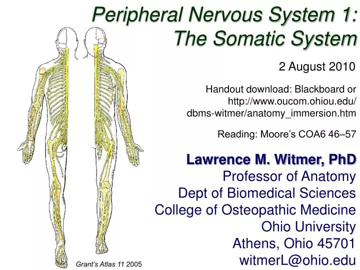
Peripheral Nervous System 1: The Somatic System. 2 August 2010. Handout download: Blackboard or http://www.oucom.ohiou.edu/ dbms-witmer/anatomy_immersion.htm. Reading: Moore’s COA6 46–57. Lawrence M. Witmer, PhD Professor of Anatomy Dept of Biomedical Sciences

E N D
Peripheral Nervous System 1: The Somatic System 2 August 2010 Handout download: Blackboard or http://www.oucom.ohiou.edu/ dbms-witmer/anatomy_immersion.htm Reading: Moore’s COA6 46–57 Lawrence M. Witmer, PhD Professor of Anatomy Dept of Biomedical Sciences College of Osteopathic Medicine Ohio University Athens, Ohio 45701 witmerL@ohio.edu Grant’s Atlas 11 2005
neuron glial cell Dichotomies 1. Tissues: neurons vs. glia 2. Position: CNS vs. PNS 3. Function 1: sensory vs. motor 4. Function 2: somatic vs. visceral Gray’s Anatomy 38 1999
Neurons • Dendrites: carry nerve impulses toward cell body • Axon: carries impulses away from cell body • Synapses: site of communication between neurons using chemical neurotransmitters • Myelin & myelin sheath: lipoprotein covering produced by glial cells (e.g., Schwann cells in PNS) that increases axonal conduction velocity • Demyelinating diseases: e.g., Multiple Sclerosis (MS) in CNS or Guillain- Barré Syndrome in PNS dendrites cell body axon with myelin sheath Schwann cell synapses Moore’s COA5 2006
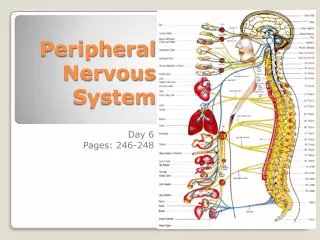
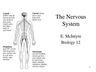
CNS vs. PNS Central Nervous System • brain & spinal cord • integration of info passing to & from the periphery Peripheral Nervous System • 12 cranial nerves • 31 pairs of spinal nerves • Naming convention changes at C7/T1 Collection of nerve cell bodies: • CNS: nucleus • PNS: ganglion Moore’s COA5 2006
Sensory (Afferent) vs. Motor (Efferent) sensory (afferent) nerve CNS e.g., skin (pseudo-) unipolar neurons conducting impulses from sensory organs to the CNS motor (efferent) nerve CNS e.g., muscle multipolar neurons conducting impulses from the CNS to effector organs (muscles & glands) Gray’s Anatomy 38 1999
Somatic vs. Visceral Langman’s Embryo 9 2004
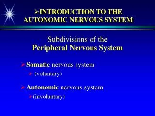
Sensory/Motor + Somatic/Visceral Somatic Nervous System Autonomic Nervous System (today) (Aug 16)
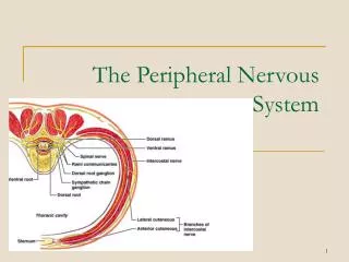
Structure of the Spinal Cord gray matter (cell bodies) • dorsal (posterior) horn • ventral (anterior) horn white matter (axons) ventral rootlets dorsal rootlets meninges pia • arachnoid • dura • denticulate ligament dorsal root (spinal) ganglion subarachnoid space (CSF) • dura • arachnoid • pia meninges spinal nerve • dorsal primary ramus • ventral primary ramus ventral root Moore’s COA5 2006
Rootlet Damage Upper brachial plexus injuries Upper Brachial Plexus Injuries • Increase in angle between neck & shoulder • Traction (stretching or avulsion) of upper rootlets (e.g., C5,C6) • Produces Erb’s Palsy Lower Brachial Plexus Injuries • Excessive upward pull of limb • Traction (stretching or avulsion) of lower rootlets (e.g., C8, T1) • Produces Klumpke’s Palsy Lower brachial plexus injuries http://www.oucom.ohiou.edu/dbms-witmer/ Downloads/2003-09-17_Ortho_Anat.pdf “Obstetrical” or “Birth palsy” • Becoming increasingly rare • Categorized on basis of damage • Type I: Upper (C5,6), Erb’s • Type II: All (C5-T1), both palsies • Type III: Lower (C8, T1), Klumpke’s Palsy Moore’s COA5 2006
Structure of Spinal Nerves: Somatic Pathways dorsal ramus dorsal root ganglion dorsal root spinal nerve somatic sensory nerve (GSA) dorsal horn CNS inter- neuron somatic motor nerve (GSE) ventral horn ventral ramus ventral root white ramus communicans Mixed Spinal Nerve sympathetic ganglion gray ramus communicans
Structure of Spinal Nerves: Somatic Pathways dorsal ramus dorsal root ganglion dorsal root spinal nerve somatic sensory nerve (GSA) dorsal horn CNS inter- neuron somatic motor nerve (GSE) ventral horn ventral ramus Somatic sensations • touch, pain, temperature, pressure • proprioception: joints, muscles Somatic motor activity: innervate skeletal muscles ventral root white ramus communicans Mixed Spinal Nerve sympathetic ganglion gray ramus communicans
Structure of Spinal Nerves: Dorsal & Ventral Rami dorsal ramus spinal nerve somatic sensory nerve (GSA) somatic motor nerve (GSE) ventral ramus Territory of Dorsal Rami (everything else, but head, innervated by ventral rami) Stern Essentials of Gross Anatomy
Impact of Lesions Disruption of sensory (afferent) neurons (paresthesia) somatic sensory nerve (GSA) somatic motor nerve (GSE)
Impact of Lesions somatic sensory nerve (GSA) somatic motor nerve (GSE) Disruption of motor (efferent) neurons (paralysis)
Impact of Lesions Disruption of sensory (afferent) neurons (paresthesia) somatic sensory nerve (GSA) somatic motor nerve (GSE) Disruption of motor (efferent) neurons (paralysis)
Impact of Lesions Disruption of sensory (afferent) neurons (back paresthesia) somatic sensory nerve (GSA) somatic motor nerve (GSE) Disruption of motor (efferent) neurons (paralysis of deep back muscles)
Segmental Innervation: Dermatomes & Myotomes somatic sensory nerve (GSA) somatic motor nerve (GSE) spinal nerve Dermatome: cutaneous (skin) sensory territory of a single spinal nerve Myotome: mass of muscle innervated by a single spinal nerve skin (dermatome) muscle (myotome) Moore’s COA5 2006
Segmental Innervation: Dermatome Maps • Based on clinical findings of deficits in cutaneous sensation • Diagnostic aids: localization of lesions to cord levels • Limits to specificity due to overlap of dermatomes dermatome overlap Moore’s COA5 2006
Dermatomes & Herpes Zoster (“Shingles)” dorsal root ganglion • Chicken pox virus (varicella) infects dorsal root ganglia • Once activated, travels along afferent axons to skin where it forms very painful rash • Often has a typical dermatomal presentation
Segmental Innervation: Myotome Maps FLEXION ABDUCTION ROTATION • Particular functions are linked to muscles innervated by particular cord levels FLEXION • Example: C5 lesion • Weakness in flexion of elbow & shoulder • Weakness in abduction & lateral rotation of shoulder Grant’s Atlas 11 2005
PNS Plexus Formation cervical plexus C1–C5 • Dermatomes: single spinal nerve • Peripheral nerves: multiple spinal nerves from different cord levels • Plexus formation: mixing of nerves from different cord levels by union and division of bundles brachial plexus C5–T1 dermatome map disparity lumbar plexus L1–L4 sacral plexus L4–S4 map of named peripheral nerves Moore’s COA5 2006
PNS Plexus Formation Example of named peripheral nerve Radial nerve receives fibers from spinal nerves from five different cord levels — in fact, all cord levels of the brachial plexus Radial Nerve C5–T1 Brachial Plexus (C5–T1) Moore’s COA5 2006
PNS Plexus Formation ABDUCT & LAT. ROTATE ABDUCT & LAT. ROTATE • Distribution of a single spinal throughout a plexus • Myotome — return to the C5 lesion example FLEX Abduction:supraspinatus & deltoid Lateral Rotation:infraspinatus & teres minor Flexion: Biceps brachii & Brachialis Moore’s COA5 2006
References Agur, A. M. R. and A. F. Dalley. 2005. Grant’s Atlas of Anatomy, 11th Edition. Lippincott, Williams & Wilkins, New York. Bannister, L. H. et al. 1999. Gray’s Anatomy, 38th Edition. Churchill Livingstone, New York. Moore, K. L. and A. F. Dalley. 2006. Clinically Oriented Anatomy, 5th Edition. Lippincott, Williams & Wilkins, New York. Sadler, T. W. 2004. Langman’s Medical Embryology, 9th Edition. Lippincott, Williams & Wilkins, New York. Stern, J. T., Jr. 1988. Essentials of Gross Anatomy. Davis, Philadelphia.