Download
1 / 17
170 likes | 185 Views
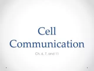
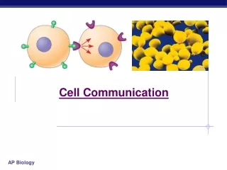
Chapter 11 (p.206-225). Cell Communication. 1. 2. 3. factor. Receptor. Figure 11.2. Exchange of mating factors. a. . a factor. Yeast cell, mating type a. Yeast cell, mating type . Mating. a. . New a/ cell. a/ . 1. 2. 3. Figure 11.3. Individual rod-shaped cells.

E N D
Chapter 11 (p.206-225) Cell Communication
1 2 3 factor Receptor Figure 11.2 Exchange of mating factors a a factor Yeast cell, mating type a Yeast cell, mating type Mating a New a/ cell a/
1 2 3 Figure 11.3 Individualrod-shapedcells Aggregation in progress 0.5 mm Spore-formingstructure(fruiting body) 2.5 mm Fruiting bodies
Plasma membranes Figure 11.4 Gap junctionsbetween animal cells Plasmodesmatabetween plant cells (a) Cell junctions (b) Cell-cell recognition
Figure 11.5a Local signaling Electrical signalalong nerve celltriggers release ofneurotransmitter. Target cell Neurotransmitter diffuses across synapse. Secretingcell Secretoryvesicle Local regulatordiffuses throughextracellular fluid. Target cellis stimulated. (b) Synaptic signaling (a) Paracrine signaling
Long-distance signaling Figure 11.5b Endocrine cell Bloodvessel Hormone travelsin bloodstream. Target cellspecificallybinds hormone. (c) Endocrine (hormonal) signaling
2 1 3 Figure 11.7d Gate closed Gate closed Ions Gate open Plasmamembrane Cellularresponse
1 2 4 3 Figure 11.7b G protein-coupledreceptor Plasmamembrane Activatedreceptor Signalingmolecule Inactiveenzyme GTP GDP GDP CYTOPLASM Enzyme G protein(inactive) GTP GDP Activatedenzyme GTP GDP P i Cellular response
2 1 3 4 Signalingmolecule (ligand) Ligand-binding site Figure 11.7c Signalingmolecule Tyr Tyr Tyr Tyr Tyr Tyrosines Tyr Tyr Tyr Tyr Tyr Tyr Tyr Tyr Tyr Tyr Tyr Tyr Tyr CYTOPLASM Receptor tyrosinekinase proteins(inactive monomers) Dimer Activated relayproteins Cellularresponse 1 P Tyr P Tyr P Tyr Tyr Tyr Tyr P Tyr P Tyr P P Tyr Tyr Tyr Tyr P Cellularresponse 2 Tyr P Tyr P Tyr Tyr P Tyr Tyr P 6 ADP 6 ATP Inactiverelay proteins
EXTRACELLULARFLUID Hormone(testosterone) Figure 11.9-5 Plasmamembrane Receptorprotein Hormone-receptorcomplex DNA mRNA NUCLEUS New protein CYTOPLASM
Figure 11.10 Inactiveprotein kinase1 Activeprotein kinase1 Inactiveprotein kinase2 ATP ADP P Activeprotein kinase2 PP P i Inactiveprotein kinase3 ATP ADP P Activeprotein kinase3 PP P i ATP P ADP PP P i
Figure 11.11 Adenylyl cyclase Phosphodiesterase H2O P i P ATP cAMP AMP
Figure 11.12 G protein GTP G protein-coupledreceptor ATP cAMP Proteinkinase A
5 4 3 2 1 RESULTS Figure 11.17 Wild type (with shmoos) formin Fus3 CONCLUSION G protein-coupledreceptor Formin P Fus3 Actinsubunit GTP P GDP Formin Formin P Microfilament Fus3 Fus3 P
Figure 11.21 Mitochondrion ActiveCed-4 ActiveCed-3 Otherproteases Ced-4 Ced-3 Nucleases (b) Death signal (a) No death signal
Figure 11.22 Cells undergoingapoptosis Space betweendigits 1 mm Interdigital tissue