Download

1 / 30
300 likes | 419 Views
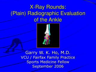
X-Ray Rounds. Wrist Mark Scott Nov. 8, 2007. Systematic Approach. Fracture Identification, Look for: Mal-alignment Discontinuity Radio-lucency / radio-opacity Fat pads. 11-22-11 Rule. Radius tilted ~11 0 volar on Lat. Radius tilted ~22 0 ulnar on AP

E N D
X-Ray Rounds Wrist Mark Scott Nov. 8, 2007
Systematic Approach Fracture Identification, Look for: • Mal-alignment • Discontinuity • Radio-lucency / radio-opacity • Fat pads
11-22-11 Rule • Radius tilted ~110 volar on Lat. • Radius tilted ~220 ulnar on AP • Radial Styloid ~11mm distal to ulna
11mm 22
11 22
3 C’s Rule • Distal Radius Lunate Capitate appears as 3 C’s on lateral
Distal Radial # • Apply 11-22-11 rule • Ortho referral for open, comminuted, unstable or failure to reduce, DRUJ, and NV compromise3 • Generally, Smith # (volar angl.) more unstable than Colles # • Research study4
22 Dorsal Barton’s Fracture
Barton’s Fracture • Intra-articular Shearing injury of dorsal (or volar) radial lip. • Require ortho referral due to high-degree of instability (insertion of Brachioradialis tendon)
Scaphoid # • Most commonly # carpal bone (60-70%) • Axial loading 70-100% sensitive (better than snuff box tenderness) • Evidence suggests below elbow cast with neutral wrist & thumb free is adequate3 • Refer if >1mm displaced or comminuted • Follow up within 1 week is crucial.
CT vs MRI vs Bone Scan for Scaphoid # • Radiographs miss 10-20% of scaphoid # • CT more sensitive and readily available3 • MRI more info re: ligamentous injury but ties up MRI time. • Bone scan very sensitive (72hrs - 2 weeks) but non-specific3 • High resolution US may be imaging modality of choice in future (Sn100%, Sp98%)
Scapho-lunate Dissociation • Forceful hyper-ext of the wrist • Tenderness immediately distal to Lister’s tubercle • Terry Thomas Sign or signet ring sign • Ortho referral and look for Lunate/Perilunate dislocation
Summary • Clinical Scaphoid CT if can’t immobilize or cast & f/u bone scan in 3-5 days.4 • Gross reduction of Colles # is adequate to prevent negative sequelae.1 • Stability: Colles > Smith’s > Barton’s • Obtain multiple views and use 11-22-11 and 3 C’s rules.
References • Jaremko JL et Al. Do radiographic indices of distal radius fracture reduction predict outcomes in older adults receiving conservative treatment?Clinical Radiology. 62(1):65-72, 2007 Jan. • McRae, R. Pocketbook of orthopaedics and fractures [2nd ed.]. Churchill Livingstone Elsevier, 2006. • Ritchie JV. Emergency Emerg Med Clin North Am - 01-NOV-1999; 17(4): 823-42 • Seitz et al. Fractures and dislocations of the wrist. Rockwell and Green’s Fractures in Adults [5 ed]. Lippincott, Williams & Wilkins, 2002. • Tintnelli, JE. Emergency medicine: a comprehensive study guide [6th ed]. American College of Emergency Physicians / McGraw-Hill, New York, 2004. Pp. 1674-84.
Thank You 22