Download
1 / 82
820 likes | 938 Views
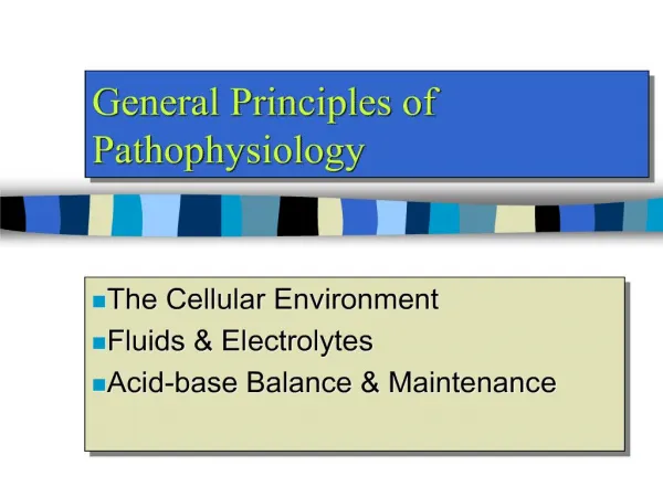
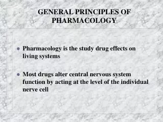
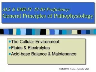
General Principles of Pathophysiology. The Cellular Environment Fluids & Electrolytes Acid-base Balance & Maintenance. Topics. Describe the distribution of water in the body Discuss common physiologic electrolytes Review mechanisms of transport osmosis, diffusion, etc

E N D
General Principles of Pathophysiology The Cellular Environment Fluids & Electrolytes Acid-base Balance & Maintenance
Topics • Describe the distribution of water in the body • Discuss common physiologic electrolytes • Review mechanisms of transport • osmosis, diffusion, etc • Discuss hemostasis & blood types • Discuss concepts of acid-base maintenance
Distribution of Water • Total Body Weight/ Total Body Water • Intracellular - ICF (45%/75%) • Extracellular - ECF (15%/25%) • Intravascular (4.5%/7.5%) • Interstitial (10.5%/17.5%)
Fluid Distribution Extracellular Intra- cellular 45% 31.5 kg Interstitial 10.5 % 7.35 kg Intra- vascular 4.5% 3.15 kg Capillary Membrane Cell Membrane Total Body Weight
Fluid Distribution Extracellular Intra- cellular 75% 31.5 L Interstitial 17.5 % 7.35 L Intra- vascular 7.5% 3.15 L Capillary Membrane Cell Membrane Total Body Water
Fluid Intake Water from metabolism: 200 ml (8%) Water from beverages: 1600 ml (64%) Water from food: 700 ml (28%)
Fluid Output Water from lungs: 300 ml (11%) Water from feces: 150 ml (5%) Water from skin: 550 ml (25%) Water from urine: 1500 ml (59%)
Osmosis is the net movement of water from an area of LOW solute concentration to an area of HIGHER solute concentration across a semi-permeable membrane. diffusion of water in terms of [water] Diffusion is the net movement of solutes from an area of HIGH solute concentration to an area of LOWER solute concentration. Osmosis versus Diffusion
Tonicity • Isotonic • Hypertonic • Hypotonic
Isotonic Solutions • Same solute concentration as RBC • If injected into vein: no net movement of fluid • Example: 0.9% sodium chloride solution • aka Normal Saline
Hypertonic Solutions • Higher solute concentration than RBC • If injected into vein: • Fluid moves INTO veins
Hypotonic Solutions • Lower solute concentration than RBC • If injected into vein: • Fluid moves OUT of veins
Affects of Hypotonic Solution on Cell • The [solute] outside the cell is lower than inside. • Water moves from low [solute] to high [solute]. • The cell swells and eventually bursts! Ruptured Cell Swollen Cell Swelling Cell Cell
Shrinking Cell Shrunken Cell Affects of Hypertonic Solution on Cell • The [solute] outside the cell is higher than inside. • Water moves from low [solute] to high [solute]. • The cell shrinks! Cell
No fluid movement • Fluid movement into veins • Fluid movement out of veins • Infusion of isotonic solution into veins • Infusion of hypertonic solution into veins • Infusion of hypotonic solution into veins
Ion Distribution Anions Cations Na+ Cl- HCo3- Extracellular Protein- PO4 - Ca+ K+ Mg+ 3 Intracellular
Example of Role of Electrolytes • Nervous System • Propagation of Action Potential • Cardiovascular System • Cardiac conduction & contraction
Composition of Blood • 8% of total body weight • Plasma: 55% • Water: 90% • Solutes: 10% • Formed elements: 45% • Platelets • Erythrocytes
Hematrocrit • % of RBC in blood • Normal: • 37% - 47% (Female) • 40% - 54% (Male)
Blood Components • Plasma: liquid portion of blood • Contains Proteins • Albumin (60%) contribute to osmotic pressure • Globulin (36%): lipid transport and antibodies • Fibrinogen (4%): blood clotting
Blood Components • Formed Elements • Erythrocytes • Leukocytes • Thrombocytes
Erythrocytes • ‘biconcave’ disc • 7-8 mcm diameter • Packed with hemoglobin • 4.5 - 6 million RBC/mm3 (males) • 120 day life span • 2 million replaced per second!
Leukocytes • Most work done in tissues • 5,000 - 6,000/mm3 • Neutrophils (60-70%) • Basophils (Mast Cells) (<1%) • Eosinophils (2-4%) • Lymphocytes (20-25%) • Monocytes (Macrophages) (3-8%)
Thrombocytes • Platelets • Cell fragments • 250,000 - 500,000/mm3 • Form platelet plugs
Hemostasis • The stoppage of bleeding. • Three methods • Vascular constriction • Platelet plug formation • Coagulation
Coagulation • Formation of blood clots • Prothrombin activator • Prothrombin Thrombin • Fibrinogen Fibrin • Clot retraction
Coagulation Prothrombin Activator Clot Prothrombin Thrombin Fibrinogen Fibrin
Fibrinolysis • Plasminogen • tissue plasminogen activator (tPA) • Plasmin
Blood Types • Agglutinogens (Blood Antigens) • Agglutinins (Blood Antibodies) • Agglutination (RBC clumping) • ABO • Rh Antigens
Capillary Network • Blood enters capillary network from arterioles • Flows through capillary network into venules • Arteriolar capillaries • Venous capillaries • True capillaries • Thoroughfare channels • Capillary sphincters
Sympathetic Innervation • Sympathetic fibers innervate all blood vessels except: • Capillaries • Capillary sphincters • Most metarterioles • Vasoconstrictor and vasodilator fibers
Diffusion across Capillary Wall • Capillary flow • Hydrostatic pressure • Osmotic pressure • Oncotic pressure • Capillary and membrane permeability
Edema • Fluid accumulation in the interstitial compartment • Causes: • Lymphatic ‘leakage’ • Excessive hydrostatic pressure • Inadequate osmotic pressure
Alterations in Water Movement • Edema • Fluid accumulation in interstitial spaces • Due to any condition that leads to: • Net movement of fluid out of capillaries into interstitial tissues
Pathophysiology of Edema • Normal interstitial space fluid depends on: • Capillary hydrostatic pressure • Oncotic pressure by blood plasma proteins • Capillary permeability • Lymphatic channels collect fluid forced from capillaries by blood hydrostatic pressure and return it to circulation
Mechanisms Responsible for Edema • Increased hydrostatic pressure • Decreased plasma oncotic pressure • Increased capillary permeability • Lymphatic obstruction • Increased capillary hydrostatic pressure • Venous obstruction • Sodium and water retention
Electrolyte Imbalances • In addition to water and sodium imbalances, other electrolyte imbalances may occur • Potassium • Calcium • Magnesium
Potassium • Major intracellular cation • Needed for nerve, cardiac, skeletal function • Excess excreted by kidneys • Imbalance can cause sudden death
Hypokalemia • Poor absorption, vomiting, diarrhea, renal disease, diuretics • Malaise, weakness, dysrhythmias, decreased reflexes, faint heart sounds, hypotension, anorexia, vomiting • Hospital treatment • Oral or IV potassium
Hyperkalemia • Renal failure, burns, crush injuries, infections, excessive use, acidosis • Dysrhythmias, irritability, abdominal distention, nausea, diarrhea, oliguria, weakness, paralysis • Treatment • Life threats – calcium, glucose, insulin IV, albuterol • Hospital – K+ restriction, exchange resins, dialysis
Calcium • Essential for: • Neuromuscular transmission • Cell membrane permeability • Hormone secretion • Bone growth • Muscle contraction