Download
1 / 26
370 likes | 855 Views
Tissue Types Overview. Tissue Definitions Epithelial Tissue Simple and Stratified Connective Tissue Characteristics Bone, Cartilage, Dense Connective, Loose Connective Blood Muscle Tissue Skeletal, Cardiac, and Smooth Nervous Tissue Tissue Repair Tissue Development and Aging.

E N D
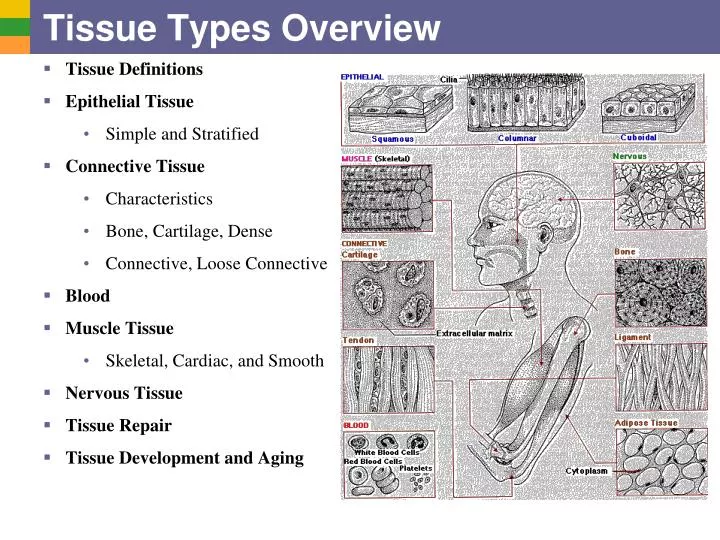
Tissue Types Overview • Tissue Definitions • Epithelial Tissue • Simple and Stratified • Connective Tissue • Characteristics • Bone, Cartilage, Dense • Connective, Loose Connective • Blood • Muscle Tissue • Skeletal, Cardiac, and Smooth • Nervous Tissue • Tissue Repair • Tissue Development and Aging
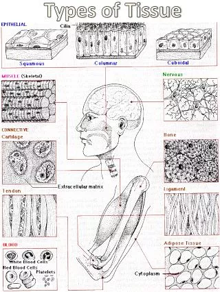
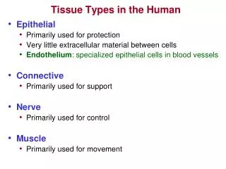
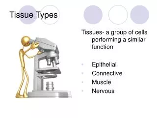
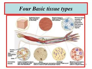
Body Tissues • Cells are specialized for particular functions • Tissues • Groups of cells with similar structure and function • Four primary types • Epithelium • Connective tissue • Muscle • Nervous tissue
Epithelial Tissues • Found in different areas • Body coverings • Body linings • Glandular tissue • Functions • Protection • Absorption • Filtration • Secretion
Epithelium Characteristics • Cells fit closely together • Tissue layer always has one free surface • The lower surface is bounded by a basement membrane • Avascular (have no blood supply) • Regenerate easily if well nourished
(a) Simple squamous epithelium Description: Single layer of flattened cells with disc-shaped central nuclei and sparse cytoplasm; the simplest of the epithelia. Air sacs of lung tissue Function: Allows passage of materials by diffusion and filtration in sites where protection is not important; secretes lubricating substances in serosae. Nuclei of squamous epithelial cells Location: Kidney glomeruli; air sacs of lungs; lining of heart, blood vessels, and lymphatic vessels; lining of ventral body cavity (serosae). Photomicrograph: Simple squamous epithelium forming part of the alveolar (air sac) walls (125x). Other important locations: Endothelium- the lining of lymphatic vessels, blood vessels, and heart Mesothelium- the epithelium of serous membranes in the ventral body cavity
(b) Simple cuboidal epithelium Description: Single layer of cubelike cells with large, spherical central nuclei. Simple cuboidal epithelial cells Function: Secretion and absorption. Basement membrane Location: Kidney tubules; ducts and secretory portions of small glands; ovary surface. Connective tissue Photomicrograph: Simple cuboidal epithelium in kidney tubules (430x). Figure 4.3b
(c) Simple columnar epithelium Description: Single layer of tall cells with round to oval nuclei; some cells bear cilia; layer may contain mucus- secreting unicellular glands (goblet cells). Simple columnar epithelial cell Function: Absorption; secretion of mucus, enzymes, and other substances; ciliated type propels mucus (or reproductive cells) by ciliary action. Location: Nonciliated type lines most of the digestive tract (stomach to anal canal), gallbladder, and excretory ducts of some glands; ciliated variety lines small bronchi, uterine tubes, and some regions of the uterus. Basement membrane Photomicrograph: Simple columnar epithelium of the stomach mucosa (860X). Figure 4.3c
(d) Pseudostratified columnar epithelium Description: Single layer of cells of differing heights, some not reaching the free surface; nuclei seen at different levels; may contain mucus- secreting cells and bear cilia. Cilia Mucus of mucous cell Pseudo- stratified epithelial layer Function: Secretion, particularly of mucus; propulsion of mucus by ciliary action. Location: Nonciliated type in male’s sperm-carrying ducts and ducts of large glands; ciliated variety lines the trachea, most of the upper respiratory tract. Basement membrane Photomicrograph: Pseudostratified ciliated columnar epithelium lining the human trachea (570x). Trachea Figure 4.3d
(e) Stratified squamous epithelium Description: Thick membrane composed of several cell layers; basal cells are cuboidal or columnar and metabolically active; surface cells are flattened (squamous); in the keratinized type, the surface cells are full of keratin and dead; basal cells are active in mitosis and produce the cells of the more superficial layers. Stratified squamous epithelium Function: Protects underlying tissues in areas subjected to abrasion. Nuclei Location: Nonkeratinized type forms the moist linings of the esophagus, mouth, and vagina; keratinized variety forms the epidermis of the skin, a dry membrane. Basement membrane Connective tissue Photomicrograph: Stratified squamous epithelium lining the esophagus (285x). Figure 4.3e
Stratified Epithelium, Other Types • Stratified cuboidal • Stratified columnar • Quite rare in body • Found in some sweat and mammary glands • Typically two cell layers thick • Limited distribution in body • Small amounts in pharynx, male urethra, and lining some glandular ducts • Also occurs at transition areas between two other types of epithelia Mucous gland duct, tongue
(f) Transitional epithelium Description: Resembles both stratified squamous and stratified cuboidal; basal cells cuboidal or columnar; surface cells dome shaped or squamouslike, depending on degree of organ stretch. Transitional epithelium Function: Stretches readily and permits distension of urinary organ by contained urine. Location: Lines the ureters, urinary bladder, and part of the urethra. Basement membrane Connective tissue Photomicrograph: Transitional epithelium lining the urinary bladder, relaxed state (360X); note the bulbous, or rounded, appearance of the cells at the surface; these cells flatten and become elongated when the bladder is filled with urine. Figure 4.3f
Glandular Epithelium • Gland – one or more cells that secretes a particular product • Two major gland types • Endocrine gland • Ductless • Secretions are hormones • Exocrine gland • Empty through ducts to the epithelial surface • Include sweat and oil glands • Found as unicellular and multicellular types
Unicellular Exocrine Glands: Goblet Cell • The only important unicellular gland is the goblet cell Microvilli Secretory vesicles containing mucin Rough ER Golgi apparatus Nucleus (a) (b) Figure 4.4
Multicellular Exocrine Glands • Multicellular exocrine glands are composed of a duct and a secretory unit • Classified according to: • Duct type (simple or compound) • Structure of their secretory units (tubular, alveolar, or tubuloalveolar)
Simple duct structure (duct does not branch) Compound duct structure (duct branches) Tubular secretory structure Simple tubular Simple branched tubular Compound tubular Example Intestinal glands Example Stomach (gastric) glands Example Duodenal glands of small intestine Alveolar secretory structure Simple alveolar Simple branched alveolar Compound alveolar Compound tubuloalveolar Example No important example in humans Example Sebaceous (oil) glands Example Mammary glands Example Salivary glands Surface epithelium Duct Secretory epithelium Figure 4.5
Modes of Secretion • Merocrine • Products are secreted by exocytosis (e.g., pancreas, sweat and salivary glands) • Holocrine • Products are secreted by rupture of gland cells (e.g., sebaceous glands)
Tissue Types Overview • Tissue Definitions • Epithelial Tissue • Simple and Stratified • Connective Tissue • Characteristics • Bone, Cartilage, Dense • Connective, Loose Connective • Blood • Muscle Tissue • Skeletal, Cardiac, and Smooth • Nervous Tissue • Tissue Repair • Tissue Development and Aging
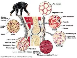
Connective Tissue • Found everywhere in the body • Includes the most abundant and widely distributed tissues • Functions • Binds body tissues together • Supports the body • Provides protection
Connective Tissue Characteristics • Variations in blood supply • Some tissue types are well vascularized • Some have poor blood supply or are avascular • Extracellular matrix • Non-living material that surrounds living cells
Extracellular Matrix • Two main elements • Ground substance - Gel like, high water content • Proteoglycans • Glycosaminoglycans (GAGs like hyaluronan), chondroitin sulfate as dietary supplement for osteoarthritis • Glycoproteins • Fibers • Produced by the cells • Three types • Collagen fibers • Elastic fibers • Reticular fibers Reticular fibers
Connective Tissue Types: Areolar • Areolar connective tissue Generic organ packaging tissue, soaks up water
Connective Tissue Types: Bone • Bone (osseous tissue) Has both an organic and a mineral component
Tissue Types Overview • Tissue Definitions • Epithelial Tissue • Simple and Stratified • Connective Tissue • Characteristics • Bone, Cartilage, Dense • Connective, Loose Connective • Blood • Muscle Tissue • Skeletal, Cardiac, and Smooth • Nervous Tissue • Tissue Repair • Tissue Development and Aging