Download
1 / 36
390 likes | 620 Views
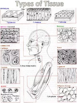
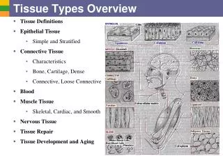
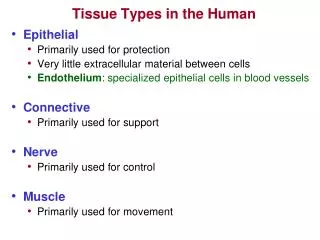
4 Tissue Types in the Body. Epithelia = epidermis of skin, lining of tubes, glands Functions in protection, absorption, secretion Muscle = functions in providing movements within the body; contractile Nervous = specialized for impulse conduction, serves as long-range communication system

E N D
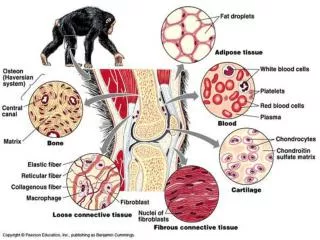
4 Tissue Types in the Body • Epithelia = epidermis of skin, lining of tubes, glands • Functions in protection, absorption, secretion • Muscle = functions in providing movements within the body; contractile • Nervous = specialized for impulse conduction, serves as long-range communication system • Connective Tissue = functions in support, binding and protection of soft tissues; 2 general types … • CT Proper = binding function; includes tendons, ligaments, fascia • CT Supportive = support function; includes skeletal elements – cartilage and bone
Connective Tissue Characteristics • All CT contain … • Cells – many types, generally widely spaced • Fibers (protein) = collagen, elastic, reticular • Ground substance = glycoprotein, gel-like intercellular material • All CT embryonically derived from mesenchyme • Mesenchyme develops from mesoderm, or in a few cases, neural crest tissue
Fig 5.19 – CT Types CT Supportive
Endoskeleton vs. Exoskeleton • Exoskeletons common in invertebrate animals, often formed from proteins (e.g., arthropods) or calcium-carbonate (e.g., molluscs) • In vertebrates, exoskeleton is limited to scales and dermal bones • Both are derivatives of skin • Dermal bones from dermatome portion of epimere (somite) • Exoskeleton becomes directly associated with endoskeleton (i.e., internal), although some early fishes retain external dermal bony armor (e.g., Ostracoderms, Placoderms).
Armor composed of dermal bone covers head region of many Ostracoderms (left) and Placoderms (below)
Endoskeleton • Includes notochord, cartilage and bone • Notochord = present in all Chordates, at least in embryonic stage. • Largely replaced by vertebral column in all vertebrates except the most primitive (e.g., cyclostomes) • Recall that the hagfish lacks any vertebral development and retains an essentially complete notochord in the adult • Notochord composed of a distinctive, fluid-filled, vesicular CT, surrounded by an external fibrous sheath and a CT membrane
Notochord • Evolutionary Trend = from primitive to advanced vertebrates, notochord progressively replaced by vertebrae • Cyclostomes = present in adult as soft, flexible rod under neural tube. Little vertebral development in lampreys, none in hagfish • Elasmobranchs = shows alternate constrictions and expansions within and between vertebrae, but is complete • This condition applies to most fishes, Labyrinthodonts and some reptiles • Tetrapods = in most adults persists only as components of intervertebral discs between vertebrae
Fig 8.4 – Relationship between notochord and vertebral development in vertebrates
Cartilage • Present as the major skeletal element of embryonic vertebrates • Composed of CT with rubbery intercellular ground substance (glycoproteins, glycosaminoglycans) • Persists as adult cartilagenous skeleton only in cyclostomes, Chondrichthyes, and a few Osteichthyes • Some cartilage present in adults of all vertebrate groups, but in most advanced vertebrates plays only a minor role
Bone • Dominant skeletal material of most adult vertebrates. • Ground substance composed of crystalline calcium phosphate in which cells are embedded. • Calcium present as hydroxyapatite (calcium phosphate) crystals = 3Ca3[PO4]2Ca[OH]2
Bone Development • Intramembranous (Direct) = bone arises directly from mesenchyme. • Applies to dermal bones, generally produces spongy bone (although these may fill in to form compact bone) • Also produces sesamoid bones (= bones that develop within a tendon – patella, pisiform) • Endochondral(Cartilage Replacement) = cartilage develops from mesenchyme to form “model.” Bone replaces cartilage model. • Applies to the majority of skeletal elements and mostly produces compact bone
Fig 5.25 – Intramembranous bone formation
Fig 5.24 – Endochondral bone formation
Bone Development • Embryonically, bone is derived almost entirely from epimere (somite), although a few elements from neural crest. • Majority of bone derived from sclerotome. • Dermal bones derived from dermatome. • In primitive vertebrates, the myotome is the dominant part of the somite, sclerotome small and inconspicuous. • In amniotes sclerotome becomes much larger.
Amniote Scaal and Wiegreffe (2006). Schematic representation of an amniote (a) and anamniote (b) somite. On the left; an early stage of somite development, showing the epithelial somite prior to overt morphological compartmental-ization. On the right; a mature somite showing distinct somite compartments. In red;myotomal cells. In blue; dermatomalprecursor cells or dermatomalcells, respectively. In green;sclerotomal precursor cells or sclerotomal cells, respectively. aThe amniote epithelial somite (left) is divided into a dorsal half destined to form the dermomyotome, and a ventral half which will give rise to the sclerotome. bThe anamniote epithelial somite (left) is divided into a medial myotome and a lateral wall, which is thought to give rise to dermis and possibly muscle in later stages. It is likely to be homologous to the amniotedermatome. In the mature somite (right) the myotomal cells have differentiated to muscle fibers/lamellae in situ, the dermatomalcells have deepithelialized, and the sclerotome has formed an epithelial bud starting to form mesenchyme. Dermatome Sclerotome Myotome Anamniote
Bone Organization • Orientation of collagen fibers and ordered placement of bone cells within bony matrix is useful means of classifying bone • Nonlamellar bone (= fibro-lamellar bone) = characterized by irregular arrangement of collagen fibers in matrix • Occurs in fast-growing bone • Lamellar bone = regular arrangement of collagen fibers in matrix, accompanied by regular orientation of bone cells. • Occurs in slow-growing bone • Lines of Arrested Growth = regions in bone where growth ceases • Often due to seasonal activity (interruptions in growth due to environmental stress – cold weather for ectotherms) • More common in ectotherms, but also occurs in some endotherms
Bone Organization • Haversian bone = arrangement of bone into Haversian systems or osteons • Osteons consist of concentric rings of bone surrounding a central Haversian canal with bone cells between rings • Haversian canal contains blood vessels and canaliculi connect bone cells to Haversian canal so that cells can receive oxygen and nutrients • Volkman’s canals run perpendicular to Haversian canals to bring in blood vessels from outside the bone
Fig 5.22 – General Bone Types. (a) non-lamellar (fibro-lamellar) bone from young alligator, (b) lamellar bone from turtles (note growth rings); (c) Haversian bone (= specialized form of lamellar bone)
Dinosaurs: Ectotherms or EndothermsThe Evidence from Bone Histology • See Box Essays 3.4 and 3.6 • Bone Histology was a primary source of evidence in initial arguments in 1980s and 1990s that dinosaurs were endothermic • Argument: microarchitecture of dinosaur fossil bones lacked regular growth rings typical of ectotherms in seasonal environments → similar to birds and mammals; suggests constant Tb year-round • Suggestion = dinos were endotherms like birds and mammals with high MR and lifestyles characterized by sustained activity
Dinosaurs: Ectotherms or EndothermsThe Evidence from Bone Histology • Problems: • Many early birds (supposedly descendant from Coelurosaurian Theropod dinosaurs) had bones with growth rings, like ectotherms • Many dinos also have bone with growth rings, although fewer growth rings than in typical reptiles • Constant Tb (homeothermy) does not necessarily equate to endothermic MR • Large dinos in warm Mesozoic climates would likely have shown little Tb variation throughout the year even if ectothermic – inertial homeotherms
Erickson et al 2009 Cretaceous Bird Coelurosaur Note high degree of vascularization and woven bone structure in both species. The coelurosaur image shows a line of arrested growth (arrow).
Dinosaurs: Ectotherms or EndothermsThe Evidence from Bone Histology • Similarity in bone structure between dinosaurs and birds is likely related to fast growth rates. • Smaller dinosaurs and earliest birds show bone more typical of general reptilian bone, suggesting slower growth rates than in larger forms and slower than in modern birds. • Thus, fast growth rates are correlated with large size, not endothermy, so bone histology does not provide convincing evidence that dinosaurs were endotherms.
Classification of Skeletal Elements Dermal Components Axial Skeleton Somatic Endoskeletal Components Appendicular Visceral
Classification of Skeletal Elements • Dermal Components = arise in the skin (dermis - dermatome) via intramembranous formation. • Includes bony armor, scales (fish), elements of pectoral girdle and skull in all groups (except cyclostomes), and jaw bones (tetrapods) • Somatic Endoskeletal Components = cartilage-replacement bone from sclerotome • forms majority of internal structures • Visceral Endoskeletal Components = associated with gill arches and derivatives. • Derived from neural crest cells, so the only non-mesodermal instance of bone development. • Includes some jaw bones, hyoid apparatus, columella, bones of middle ear in mammals, bones supporting gills in fish.
Classification of Skeletal Elements • Axial Skeleton = Vertebral column and associated structures, including parts of skull, ribs, etc. • Appendicular Skeleton = limb girdles and elements of appendages
Haversian Bone L = lamellae O = osteocytes (in lacunae) C = canaliculi H = Haversian canal
Histophysiology of Bone • Intramembranous and Endochondral formation both result in identical ultrastructure – contains cells surrounded by bony matrix, but interconnected with other cells and with blood vessels by tiny canals (canaliculi). • Because of cellularity, adult bone is not a static tissue, but continuously adapts to prevailing stresses by appropriate deposition and resorption. • Also capable of repair if broken • Deposition and Resorption are under hormonal control – integrated with Calcium and Phosphate requirements within the body. • Bone serves as a storage tissue for both ions.
Fig 5.27 – Repair of broken bones. (a) blood clot forms, (b) cartilage invades break, (c) osteogenic bud invades cartilage forming woven bone, (d) remodeling produces healed bone structure
Histophysiology of Bone • Hormones controlling deposition and resorption also control blood calcium levels. • Parathyroid Hormone = promotes resorption, increases blood calcium • Calcitonin = reduces resorption, decreases blood calcium • Intense activity increases blood lactic acid levels (decreases pH) and causes dissolution of bone and calcium release. • May be important for muscular contraction.
Evolution of Skeletal Structure • Because bone is not present in any non-vertebrate chordates, it almost certainly was not present in the most primitive vertebrates. • In the earliest fossil vertebrates (ostracoderms) bone forms only a superficial armor (composed of dermal bone). There is no evidence of an ossified endoskeleton. • An ossified endoskeleton developed later than ossification of the exoskeleton, but has become the dominant feature in modern vertebrates, along with a reduction in dermal bone.
Evolution of Skeletal Structure • Why Ossify Skeleton with Calcium Phosphate (Calcium Carbonate in all Invertebrates)? • Hypothesis # 1 – Protection • Bone is harder and stronger than calcium carbonate so forms a more effective skeletal tissue. However, this seemingly is an equally good selective pressure for invertebrates – thus, it is likely that the protective function of hydroxyapatite bone is a secondary function. • So, what selective factor led to the evolution of hydroxyapatite bone instead of calcium carbonate, as in the exoskeleton of invertebrates?
Evolution of Skeletal Structure • Why Ossify Skeleton with Calcium Phosphate (Calcium Carbonate in all Invertebrates)? • Hypothesis # 2 – Metabolic Reserve with Structural Integrity • Evolution in vertebrates is characterized by increased activity levels. Increased activity leads to lactic acid buildup and to bone dissolution. • Hydroxyapatite is much more stable when exposed to lactate than is calcium carbonate. • So, evolution of hydroxyapatite bone was probably to allow a metabolic reserve of calcium and phosphate ions while still maintaining structural integrity during activity.
From Ruben and Bennett (1987, Evolution 41:1187-1197) • Implanted “silastic bones” composed of calcium carbonate(calcite) or calcium phosphate (apatite) into trout (also implanted crystals directly into peritoneum) • Exercised vs. Non-exercised (control) treatments • Exercised group showed increased loss of crystals compared to controls, and calcite showed greater reduction than apatite • Conclusion: Apatite more stable under low pH typical of exercise in vertebrates. P = peritoneal crystals E = silastic “bones” A =apatite (calcium phosphate) C = calcite (calcium carbonate)