Download
1 / 40
400 likes | 597 Views
Cytology. I. Introduction. A. Definition. B. How to Study ?. 1. Microscopy. a. Light Microscopy ( LM ). i . Advantages. Magnification. Resolution. Depth of Field. ii. Types. Figure 6.3. b. Electron Microscopy ( EM ). i. Advantages. Magnification. Resolution.

E N D
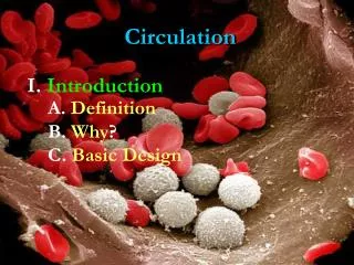
Cytology I. Introduction A. Definition B. How to Study? 1.Microscopy
a. LightMicroscopy (LM) i. Advantages Magnification Resolution Depth of Field ii. Types Figure 6.3
b. Electron Microscopy (EM) i. Advantages Magnification Resolution Depth of Field ii. Types TEM SEM Figure 6.4
a. Technique Figure 6.5 b. Advantages i. Whole samples ii. Specificity iii. Starting Point
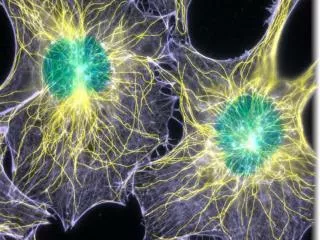
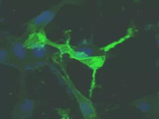
a.Vital Staining forContrast
b.Antibody Staining More Specific Contrast
II. Parts of a Cell A. Barriers 1.Cell Walls
c.Plants Figure 5.7
c.Plants Figure 6.28
1.Consistency like thickening Jell-O 2.Molecularmake-up 92% is water, 7% protein, and the rest is gases, salts, lipids, and the like dissolved in the water
Representative Animal Cell Figure 6.9
Representative Plant Cell Figure 6.9
C. Organelles = Cell Machinery 1.Membrane Bound
a.Nucleus = the keeper of the plans Envelope, pores, and nucleolus Figure 6.10
b.Endomembrane System = rER, sER, and Golgi Figure 6.12 Figure 6.13
c.House cleaners -> Lysosome or Peroxisome Figure 6.14
d.Energy Transformers= Chloroplast & Mitochondria Figure 6.17 Figure 6.18
e.Vacuoles i.Animal Types = Food or Contractile ii.Plant Types = Central, Amyloplasts, & Chromoplasts
a.Cytoskeleton Figure 6.20
b.Ribosomes c.Centriole Figure 6.11 Figure 6.22
D. Cellular Specializations 1.Microvilli 2.Cilia
Microvilli = short non-moving membrane extensions to increase cell’s overall surface area Cilia = long, moving internal cellular extensions to move something across the cell surface.
Flagella move the entire cell Figure 6.24
Flagella move the entire cell Figure 6.25
E. Intercellular Junctions 1.Plants 2.Animals
Figure 6.28 Plants Figure 4.11 Desmosomes Animals Gap Junctions Figure 6.32