Download
1 / 44
460 likes | 645 Views
PHM142 Metabolic Biochemistry and Immunology II WHITE CELLS CHEMICAL WARFARE lecture outline. Molecular mechanisms of leukocyte function Rheumatoid arthritis CGD disease 2) Eosinophils Macrophages Function Growth factors and cytokines But – macrophage - induced tissue injury

E N D
PHM142 Metabolic Biochemistry and Immunology II WHITE CELLS CHEMICAL WARFARE lecture outline • Molecular mechanisms of leukocyte function • Rheumatoid arthritis • CGD disease • 2) Eosinophils • Macrophages • Function • Growth factors and cytokines • But – macrophage - induced tissue injury • Multiple organ failure, ARDS, sepsis
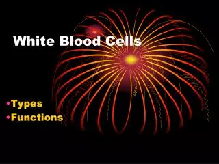
Blood Cells Main function Transport O2 and CO2 Destroy invading bacteria Phagocytose & kill invading antigens Destroy parasites and modulate allergic inflammatory reactions Release histamine, serotonin, bradykinin, heparin, and cytokines; converts arachidonic acid to prostaglandins & leukotrienes (T & B) mediate cytokine release Phagocytose & kill ingested microbes Kill virally infected cells and tumor cells (offer “natural” immunity as well as adaptive) Initiate blood clotting; also release histamine and serotonin Type of cell Red blood cells (erythrocytes) White blood cells (leukocytes) polymorphonuclear/granular leukocytes Neutrophils (50 60%) Eosinophils (1 4%) Basophils (0.5 2%) mononuclear leukocytes Lymphocytes (20 40%) Monocytes (2 9%) ( macrophages in extravascular tissue) Killer cells Megakaryocytes ( platelets)
2 1 Macrophage 3 Thin sections of the major types of white blood cells (leukocytes) found in the circulation, showing the variety of internal structures observed.
Molecular mechanisms of neutrophil function 1 Amer J Med. 109, 33-44 (2000) Neutrophil membrane electron transport CYTOSOL PHAGOSOME MEMBRANE 2 2 PHAGOSOME VACUOLE Fe2+
Inflammation or infection MPO(myeloperoxidase), HOCl(hypochlorite), SOD (superoxide dismutase). HOCl + red cell GSH = GSH cyclic sulfonamide (biomarker for HOCl formation in vivo that reflects inflammation (oxidant formed by neutrophils)) Living with a killer: The effects of hypochlorite on mammalian cells IUMB Life 50,259-266 (2000) ; Chem.Res.Toxicol.21,1011-6 (2008)
A CGD disease clinical test for the lack of leukocyte respiratory burst. Leukocyte Respiratory burst by phagosome membr. elect. transport. Leukocytes (from normal patient) Opsonized bacteria or N-formyl methionine peptide * O2 level in solution Oxygen uptake is insensitive to cyanide Time Oxygen in solution is measured by an O2 electrode superoxide detected by NBT reduction to formazan (blue precipitate). * Made by bacteria; recognized by mammalian cells as foreign. No O2 consumption by CGD patient’s leukcoytes.
Trends in Biochem. Sci. 2, 63 (1977)
Activation of leukocyte NADPH oxidase (Nox) showing assembly of the enzyme and fusion of the oxidase-containing vesicle with the phagosomal membrane. Babior, 2000. American Journal of Medicine. 109:33 44.
(phosphorylation by kinase) Proton Channel Function + H+ Proton Channel membrane b558 b558 Vacuolar space phox is “phagocyte oxidase” NADPH oxidase (Nox) activation gp91phox is b558 with proton channel (or K+?) Medicine 79, 170-200 (2000)
NADPH OXIDASE DEFICIENCY Chronic Granulomatous Disease (CGD) White Cell bact. 0.1 M Chloride in blood • CGD can kill Bacteria that produce H2O2 and have low catalase activity, e.g., streptococci. • But can’t kill bacteria which have high catalase activity or low H2O2 production, e.g., serratia, nocardia, and aspergillus; fungal, staphylococcus, burkholderia (pneumonia, sepsis).
LEUKOCYTE ELECTRON TRANSPORT Chronic granulomatous disease (CGD) • Two-thirds have mutations in gp91phox gene Unable to form H2O2. • i.e., no cyanide resistant respiratory burst & don’t live beyond childhood • Have X-linked chromosomal deficiency • Have no cyt. b558 function • b-subunit – 91kD – glycosylated – X chromosome DEFECT • a-subunit – 22kD – normal chromosome 16 • CGD patients have mutation in p47phox gene (reach adulthood) • Have autosomal recessive deficiency • CGD patients have no cytosol activating protein that phosphorylates and activates NADPH oxidase which causes translocation of the oxidase from the endoplasmic reticulum to the plasma membrane. Medicine 79, 170 200 (2000); J Leukocyte Biol. 69, 191 6, 2001
THERAPY FOR CGD DISEASE • Prophylaxis with trimethoprim-sulfamethoxazole and interferon gamma • Bone marrow transplantation or granulocyte transfusion (with severe infections) • Hemopoietic stem cell gene therapy (retrovirus-mediated expression of gp91phox to reconstitute NADPH oxidase – Dinauer et al., 1999. Blood. 94(3):914-22).. • Possible Pharmaceutical Therapy? Opsin + Chemoattractant Polystyrene Beads Glucose oxidase (Fungal) Glucose + O2 H2O2
NADPH oxidase family: tissue locationJ.Biol.Chem 283,16961-16965(2008) • NOX1 colon>prostate,uterus,breast,macrophage? • NOX2 phagocyte H2O2 for MPO >> hepatocyte,B lymphocyte, cardiomyocytes,endothelial cells • NOX3 inner ear,fetal • NOX4 kidney,blood vessels,cardiomyocytes,endoth. • NOX5 lymphoid tissue,testis • Duox1 & 2 (dual oxidase) makes thyroxine hormone and H2O2 for thyroid peroxidase TPO >lung,GI
Rheumatoid Arthritis is an autoimmune inflammatory disease that may affect (skin, blood vessels, heart, lungs, muscles-- but principally attacks joints (proliferative synovitis progressing to cartilage destruction/joint ankylosis. • Pathogenesis (unknown etiology):genetic susceptibility; joint damage mediated by leukocytes or exogenous arthritogen (virus, mycobacteria). leukocytes Synovium (synovial fluid) Babior, 2000. American Journal of Medicine. 109:33 44. From Am J Med. (2000). 190:33-44
NSAID Drug therapy for RA or OA IBUPROPHEN COX-1 inhib. 300mg 3 times pd Cardiovascular & GI bleeding risk,inhib platelets, kidney/liver tox. ALEVE (Naproxen) 200mg otc ; lasts 12h • Inhibits inflammation , COX-I and COX-2 inhibitor • Decrease pain, temp, muscle pain, menstrual cramps • Lower stroke risk than ibuprofen; high dose risk GI bleed. VIOXX COX-2 inhibitor for OA,RA 1999 introd. • 2004 (withdrawn due to heart attack,stroke). Late? CELEBREX (celecoxib)1998 COX-2 inhibitor 400mg • low cardiorenal tox, platelet effects & GI bleeding
Side effects of COX-1 inhibitors • e.g. Aspirin may cause stomach bleeding and risk GI ulcer formation. • molecular mechanism of bleeding/ulcers unknown. • Aspirin causes stomach cell mitochondrial uncoupling and acidosis. • inhibits COX-1 activity thereby increasing tissue unsat. fatty acid levels and cause acidosis. • decreases PGE2 levels that protect stomach membr. • inhibits thromboxane formn. and platelet aggreg. • unsat.fatty acid + PGS attacks protective mucous layer
Mechanism of NSAID induced GI toxicity • Stage I NSAIDs decrease intestinal mucosal prostanoids (PGE,TXB2, 6-keto-PGF) • Mitochondrial uncouplers (e.g.indomethacin,DNP) compromised intestinal barrier). Resp.inhibited • Stage 2 mild inflammation + aspirin • Stage 3 histopathology, ulcers and bleeding • Rat model: ulcers, intestinal inflammation or gastric permeability induced by indomethacin or DNP + Cox inhib (aspirin). Aliment Pharmacol. Ther. (2000) 14,639-650
Rheumatoid Arthritis Therapy Repetitive hypoxia in joints, and endothelial cells ATP hydrolysis Hypoxanthine from ATP Reperfusion O2 + endothelial cell xanthine oxidase Monosodium urate crystals in joint Uric acid ALLOPURINOLinhib. ROS endothelial cell nitric oxide Destruction of joint peroxynitrite
Anti-Inflammation therapy • ROS Scavenger Therapy SOD, selenomethionine/Vit E, 5-aminosalicylate, penicillamine:Cu • Macrophage inhibitor therapy: Gold thiomalate or auranofin or zinc or copper salicylate • Prostaglandin Synthetase Inhibitors:-(NSAIDS) COX-1 many cells e.g.aspirin,ibuprofen, but GI bleeding,kidney COX-2 inflammatory cells e.g. VIOXX (withdrawn), CELEBREX but cardiovascular problems. • “Biologics” antibodies that inhibit inflammatory cytokines e.g.TNF-α • Diet: decrease arachidonate intake (meat), increase omega 3 fatty acids (fish) decreases bad prostaglandins, decrease Fe intake References: Semin. Arthritis Rheum. 27: 366-70 (1998). Autoimmunity Reviews 7,1-7(2007) Anti-inflammatory Biologics
EOSINOPHILS: (chemical warfare cont.) • Accumulate in parasite infection, asthma, rheumatoid diseases, • Hodgkin’s lymphoma and allergic or inflammatory diseases • destroy parasitic worms, tumor cells, fungi and bacteria by forming • hypobromite • H2O2 + Br- + H+ HOBr (hypobromite) + H2O • 2) cytokine production e.g. PAF, LTC4 unlike neutrophils (leukocytes) . 2 MPO Biochem J. 358, 233-239 (2001).
MACROPHAGES - chemical warfare function 1) Endocytosis and exocytosis via specific receptors for IgG and C3 coated in bacteria 2) H2O2 production by NADPH oxidase to kill mycobacteria 3) Arachidonate oxidation to prostaglandin 4) Cytokine production - upon activation by PDGF a) lipopolysaccharide (endotoxin) TNF-a b) immune system activation BCG infection IL-1 c) inflammation or interferon (IFN-g) PAF TGF a and b arginine nitric oxide kill tumor cells 5) endocytosis and delivery to lysosomes (via scavenger receptor) of oxidised LDL (low density lipoprotein) - can result in transformation to foam cell (the basis of the formation of atherosclerotic plaque) 3
SURGERY TRAUMA e.g. if intestinal surgery causes prolonged interrupted blood flow (reperfusion injury) EARLY ORGAN FAILURE if endotoxin from Gram –ve cell wall of Gram-ve bacteria in the intestine crosses gut barrier Endotoxin releases immune cell growth factors & humoral/cellular mediators (now too late!) e.g. TNF, IL, arachidonic oxidation production Attract PMN, macrophages, endothelium cells which further release TNF,NO,H2O2 anaphylactic shock mast cells release histamine (e.g. ARDS (adult respiratory distress syndrome), hepatitis Multiorgan failure, sudden death Harrison 2007 “Principles of Medicine”
Tumor Necrosis Factor (TNFα) as the primary trigger for inflammatory response Macrophages,monocytes,lymphocytes,keratinocyte • TNFα incr in chronic inflammatory diseases: rheumatism,arthritis,encephalitis,tumors • Rheumatoid arthritis , psoriasis,Crohn’s disease • Proinflammatory > antiinflammatory cytokines • Drug therapy: NSAIDs, GC glucocorticoids, Disease Modifying Antirheumatic Drugs (DMARDs).
Drug induced hepatocyte cytotoxicity caused by activated immune cells releasing cytokines and reactive oxygen species (Kupffer cells , macrophages ,neutrophils) i 1) Toxic doses of drugs or chemicals injure hepatocytes. Injured hepatocytes release factors that attract Kupffer cells to specific regions of the liver. 2) Additional mononuclear phagocytes are also recruited from blood and bone marrow precursors. 3) Once localised in the liver, the macrophages become activated by hepatocyte-derived factors of endothelial cells. 4) Activated macrophages and endothelial cells release cytokines e.g.TNFα & platelet activating factor prime & activate Kupffer cells which release Reactive Oxygen Species and more cytokines. 5) Some chemoattractants and cytokines can attract and activate neutrophils that also contribute to hepatocyte injury.
Macrophages and Tissue Injury Toxicant Target tissues Activated macrophages e.g. Kupffer cells Mediators Gold thiomalate Gadolinium chloride (macrophage inhibitor) Amplification Cytotoxicity Tissue Injury Model for the role of macrophages in tissue injury by generating inflammatory mediators.
Macrophage killing mechanism • Macrophages endocytose & present antigens to T cells as foreign substances • Exocytose (via specific receptors) IgC and C3 coated bacteria • Produce H2O2 to kill mycobacteria • Produce cytokines PDGF,PAF,TNFα & β, IL1 convert arginine to NO • Prevents foam cell formation by endocytosis of oxid. LDL by macrophage oxid LDL & fusion with lysosomes and digestion by cathepsins & Plipases.
INFLAMMATION MODEL FOR IDIOSYNCRATIC DRUG TOXICITY (Liver injury, bone marrow agranulocytosis, lung toxicity) O’Brien et. al. Drug Discovery Today 2005 10, 617-625
Table 2: Inflammatory mediators implicated in toxicity Toxicant Mediator Lung Liver 1) Reactive oxygen intermediates Ozone Endotoxin (H2O2, .OH) Asbestos Acetaminophen Amiodarone Corynebacterium parvum Reactive nitrogen intermediates Bleomycin Galactosamine (peroxynitrite) Carbon tetrachloride 1,2-dichlorobenzene Phenobarbital Endotoxin 2) Hydrolytic enzymes Endotoxin (collagenase, elastase) Silica 3) Lipids Hyperoxia (leukotrienes, prostaglandins, thromboxanes) 4) IL-1 Cigarette smoke 5) TNF-a (mitochondrial toxin, Cadmium chloride Alcohol reactive oxygen species) Toxicology 160, 111 8 (2001). Ann. Rev. Pharmacol. Toxicol. 35, 655 (1995).
Table 1: Toxicants whose pathophysiology is thought to be associated with macrophages and inflammatory mediators Pulmonary toxicants Hepatotoxicants Ozone Acetaminophen Sulfuric acid Carbon tetrachloride Nitrogen dioxide Lipopolysaccharide Cigarette smoke Galactosamine Hyperoxia 1,2-dichlorobenzene Bleomycin Cadmium Amiodarone Allyl alcohol Titanium dioxide Endotoxin Silica Asbestos Cadmium chloride
Autoimmune disease drug therapy • 1. Alkylating Agents (anticancer cytostatic drugs) • Inhibit cell division of both T cells and B cell (lymphocytes) • Cyclophosphamide, nitrosoureas, platinum compounds • Efficient therapy for systemic lupus, autoimmune hemolytic anemias, Wegener’s • 2. Antimetabolites Interfere with synthesis of nucleic acids • a) Folic acid analogues (METHOTREXATE) binds dihydrofolate reductase and prevents synthesis of tetrahydrofolate. Used in treatment of autoimmune diseases (rheumatoid arthritis) and in transplantations • b)Purine analogues (AZATHIOPRINE, or MYCOPHENOLATE (CellCept) inhib. IMP dehydrogenase and GMP synth. Treatment of autoimmune diseases (Wegeners,psoriasis ) also prevents transplant rejection. Mercaptopurine (leukemia,Crohns). • c)Pyrimidine analogues LEFLUNOMIDE • 3. Cytotoxic anticancer e.g. mithramycin, dactinomycin inhibits RNA transcription and DNA replication
4. Biologics: anti-cytokine antibodies • TNFalfa blockers1)Etanercept for RA 2)Infliximab (remicade) for Crohns disease,colon cancer a chimeric human Fab. 3) Adalimumab (Humira) a humanised monoclonal for colon cancer,macular degen. 4)Stelera NEW for psoriasis, 5 shots p.a.$50K • Rituximab kills CD20 B cell NH lymphomas,RA • Abatacept,fusion protein inhib.T cell costim.RA • Avastin (bevacizumab) inhib.VEGF-A (vascular endoth.growth factor) glioblastoma,macular degen.
T lymphocytes formed in b.m.& mature in thymus 1) T helper cells help B cells mature into plasma cells. CD4+T cells express CD4 protein on their surface 2)19% Cytotoxic T cells destroy viral infective & tumor cells 3)Memory Tcells CD4+(lost in AIDs) orCD8+(cytotoxic) subset of antigen T cells that persist long after infection. 4) Regulatory T cells (suppressor T cells) 5)Natural killer (NK) largest T cell.Interferons cause cytotoxic granule release 6) Antigen-presenting cell (APC)
B cells bursa of fabricius(birds) • Plasma cells are large B cells exposed to antigens which produce antibodies that bind to microbes. In tissues not plasma. • Contain rough e.r. & cell rapid apoptosis (short life) • Memory B cells formed from activ.B cells(long life) • 23% B-1 cells IgM>IgG in peritoneal & pleural cavities • B-2 cells
B cell depletion therapy Immunosuppressants and anti-inflammatories RA • B cell depleting agents like rituximab • tocilizumab human monoclonal antibody against IL-6R cytokine No truly effective therapies exist
Biomarkers for inflammation disease Pro-inflammatory cytokines • Elevated CRP,IL-1B,IL-1B,IL-6,IL-8 • Tumor necrosis factor alpha • c-ANCA antineutrophil cytoplasmic stain • p-ANCA antineutrophil perinuclear stain • x-ANCA chronic inflammatory bowel disease
Biomarkers for determining oxidative stress associated with diseases • 1) Plasma lipid hydroperoxide • 2) Oxidative DNA damage DNA 8-OHdQ • Urinary 8-OHdQ • Thymine oxidation HMdU • DNA strand breaks (Comet assay) • 3) Protein carbonyls using dinitrophenylhydrazine • Protein oxidation,cysteine,methionine,histidine • Protein AGEs • 4) Fructose oxidation forms toxic glyoxal
Celiac disease (dietary autoimmune) • Small intestine inflammation caused by an immune reaction to gliadin, a gluten protein, found in wheat,barley,rye (bread). • Blunting of villi, damage to inner surface , lymphocyte infiltration of crypts. • Abdominal pain and diarrhea. • Impaired vitamin uptake (brain,nervous,bone,liver) • Treatment life long gluten free diet • 1:>105-1:750 starting middle infancy
Macrophages • White blood cells within tissue, have a role in innate and adaptive immunity • They engulf pathogens and debris via phagocytosis, and move around via amoeboid movement
The Immune System • When a pathogen (can be self in this case) is ingested by a macrophage, pathogen proteins attach it to class II MHC • Macrophage activated to deliver signals to T-cells which produces autocrines and stimulate their own production • Helper T cells activate B cells which produce antibodies that inhibit the pathogens
Autoimmune Disease: Genes • Results from a loss of immunological tolerance – which is the ability to ignore self-antigens • T and B lymphocytes that recognize self-antigens are usually destroyed in the Thymus and Bone marrow, respectively, preventing autoimmunity. • Infection and overstimulation of APCs can break tolerance and induce priming of T-cells • A combination of genetics and environment are responsible for autoimmune disease • Human Lymphocyte Antigen (HLA/MHC) is the best predictor as it enhances antigen presentation resulting in increased T-cell activation
Autoimmune Disease - Damage Cell-mediated (T cells) • Immune cells both kill cells directly and indirectly via cytokines (PG, NO, etc.) • Macrophages: • INITIATE the response as antigen presenting cells • PARTAKE in killing cells through antibody dependent-dependent cell-mediated cytotoxicity and by releasing cytokines (TNF and IL-1) • PRESENT SELF-TISSUE TO T CELLS Antibody-mediated (B cells) • Binding of antigens on the surfaces of B-cells produces antibodies • Autoantibodies: • Bind to self-tissue, and activates the complement cascade which targets the self-antigen to be phagocytosed (opsonized) by Macrophages
II White (Immune) Cells • THE END