Download
1 / 60
670 likes | 1.43k Views
The Endocrine System. Endocrine and nervous systems work together Endocrine system hormones released into the bloodstream travel throughout the body target is usually far from site of synthesis binds to receptors on or in target targets = cells throughout the body

E N D
The Endocrine System • Endocrine and nervous systems work together • Endocrine system • hormones released into the bloodstream travel throughout the body • target is usually far from site of synthesis • binds to receptors on or in target • targets = cells throughout the body • results may take hours, but last longer • Nervous system • certain parts release hormones into blood • rest releases neurotransmitters that excite or inhibit nerve, muscle & gland cells • results in milliseconds, brief duration of effects
General Functions of Hormones • Help regulate: • extracellular fluid • metabolism • biological clock • contraction of cardiac & smooth muscle • glandular secretion • some immune functions • Growth & development • Reproduction
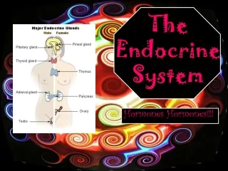
Endocrine Glands Defined • Exocrine glands • secrete products into ducts which empty into body cavities or body surface • sweat, oil, mucous, & digestive glands • Endocrine glands • secrete products (hormones) into bloodstream • pituitary, thyroid, parathyroid, adrenal, pineal • other organs secrete hormones as a 2nd function • hypothalamus, thymus, pancreas,ovaries,testes, kidneys, stomach, liver, small intestine, skin, heart & placenta
Extracellular Signaling: Mechanisms • most signals produced by cells within the body bind to receptors that are specific for that signal • most receptors are found on the cell surface • although some can be found within the cell! • binding of the signal (ligand) to the receptor results in a series of events (signal transduction) within the cell that changes the cells function • e.g. may change the transcription rate of a gene – effects protein production
Hormones: Mechanisms of Signaling • hormone producing cell = endocrine cell • e.g. thyroid, pituitary • Autocrine signaling • cell responds to the hormone it produces • Paracrine signaling • local action • local hormone (paracrine hormones) act on neighboring cells • autocrines act on same cell that secreted them • Endocrine signaling • circulating hormones (endocrine hormones) • act on distant targets • travel in blood
Types of Hormones • water-soluble • lipid -soluble
Lipid-soluble Hormones • Steroids • lipids derived from cholesterol • made in SER • different functional groups attached to core of structure provide uniqueness • e.g. cortisol, progesterone, estrogen, testosterone, aldosterone • Thyroid hormones • tyrosine ring plus attached iodines • are lipid-soluble • Retinoic acid • lipids derived from retinol (vitamin A) • regulate proliferation, differentiation and death of many cell types • some vitamins can acts a lipid-soluble hormones • e.g. vitamin D • Nitric oxide (NO) - gas testosterone aldosterone cortisol
Lipid-soluble Hormones • Eicosanoids • prostaglandins or leukotrienes • derived from arachidonic acid (fatty acid) • AA is converted either into prostaglandin H or into the leukotrienes • conversion of AA into prostaglandins is regulated by the COX enzymes • both act in the inflammatory reaction • e.g. stimulate smooth muscle cells to contract • e.g. stimulate nerve cells – pain
Water-soluble Hormones • Amine, peptide and protein hormones • modified amino acids to protein chains • serotonin, melatonin, histamine, epinephrine, insulin, dopamine • protein hormones – comprised of one or many polypeptide chains • insulin, glucagon • peptide hormones – comprised of chains of amino acids • e.g. growth hormone, oxytocin • amine hormones – derived from the amino acids tyrosine or tryptophan • epinephrine (tyrosine and phenylalanine), serotonin (tryptophan), dopamine (tyrosine) • can also act as neurotransmitters insulin
Hormone must be carried by a transport protein that allows it to dissolve within the aqueous (watery) environment of the blood plasma Hormone diffuses through phospholipid bilayer & into cell the receptor is located within the cell (cytoplasm or the nucleus) binding of H to R results in its translocation into the nucleus the H then binds directly to specific sequences within the DNA = response elements this binding turns on/off specific genes – activates or inhibits gene transcription if turned on - new mRNA is formed & directs synthesis of new proteins new protein alters cell’s activity if turned off – no new protein results and the cell’s activity is altered Action of Lipid-Soluble Hormones: Endogenous signaling
Action of Lipid-Soluble Hormones • some lipid-soluble hormone don’t cross the plasma membrane – too large • therefore they bind with receptors on the cell surface and trigger signaling events within the cells • signal similar to water-soluble hormones • e.g. prostaglandins
Action of Water-Soluble Hormones • easily travels through the blood - hydrophilic • but cannot diffuse through plasma membrane! • therefore absolutely requires the expression of receptors on the cell surface – integral membrane proteins that act as first messenger • the receptor protein activates a series of signaling events within the cells • e.g. epinephrine binds to receptor and activates an adjacent G-protein in membrane • G-protein activates adenylate cyclase to convert ATP to cyclic AMP (cAMP) in the cytosol • cAMP is the 2nd messenger • cAMP activates a series of proteins in the cytosol called kinases • kinases act to phosphorylate their targets – either activating them or inhibiting them • this speeds up/slows down physiological responses within the cell • phosphodiesterase inactivates cAMP quickly • many second messengers are made in cells in response to specific hormones • e.g. calcium, IP3, DAG • Cell response is turned off unless new hormone molecules arrive • this mechanism allows for amplification – one H-R combination can activate two G proteins which activates 4 kinases which activate 16 more kinases etc…….
Hormonal Interactions • Permissive effect • Second hormone permits the action of the first • estrogen & LH are both needed for oocyte production • Synergistic effect • two hormones act together for greater effect (vs. individually) • thyroid strengthens epinephrine’s effect upon lipolysis • Antagonistic effects • two hormones with opposite effects • insulin promotes glycogen formation & glucagon stimulates glycogen breakdown
Control of Hormone Secretion • Regulated by signals from nervous system, chemical changes in the blood or by other hormones • Negative feedback control (most common) • decrease/increase in blood level is reversed • Positive feedback control • the change produced by the hormone causes more hormone to be released • Disorders involve either hyposecretion or hypersecretion of a hormone
Hypothalamus and Pituitary Gland • Both are master endocrine glands since their hormones control other endocrine glands • Hypothalamus is a section of brain above where pituitary gland is suspended from stalk • Hypothalamus receives input from cortex, thalamus, limbic system & internal organs • Hypothalamus controls pituitary gland with 7 different releasing & inhibiting hormones – PLUS two additional hormones (ADH & oxytocin)
hypothalamus • -Emotions, autonomic functions, hormone production • -mamillary bodies – serve as relay stations for reflexes related to smell • -supraoptic and preoptic regions that function in vision • -major functions: • 1. control of the ANS – integrates signals from the ANS (regulated smooth and cardiac muscle contraction) • major regulator of visceral activities (heart rate, food movements, contraction of bladder) • 2. produces hormones & connects with pituitary to regulate its activity 3. regulates emotional and behavioral patterns – rage, aggression, pain and pleasure + sexual arousal 4. regulates eating & drinking – hypothalamus contains a thirst center which responds to a rise in osmotic pressure in the ECF (dehydration) 5. controls body temperature – monitors temp of blood flowing through the hypothalamus
median eminence Pituitary Gland • cells secrete 9 different hormones • regulates the activity of the pituitary through its secretion of 7 specific releasing and inhibiting hormones • e.g. pituitary hormone = somatotropin • hypothalmic hormones = somatotropin releasing hormone (SRH) and somatotropin inhibiting hormone (SIH) • also secretes two specific hormones that are stored in the pituitary – oxytocin and anti-diuretic hormone (ADH) • portion that marks the connection with the pituitary = median eminence • near the ME is a group of cells = neurosecretory cells • secrete the hypothalmic releasing and inhibiting hormones that regulate the pituitary • stored in synaptic vesicles • released upon production of an action potential via exocytosis into the blood stream • Diffusion down to the anterior pituitary
-hypothalamic hormones (releasing hormones) reach the pituitary via a portal system of capillaries = hypophyseal portal system (HPS) -flow of blood from the hypothal. into portal veins that carry blood to the capillaries of the ant pituitary -superior hypophyseal arteries are branches off of the internal carotid- -bring blood into the hypothal. -where the hypothal. and infundibulum meet they divide into the primary plexus of the HPS -from this plexus blood drains into hypophyseal portal veins -at the anterior pituitary these portal veins divide into the secondary plexus of the HPS -release of the hypothalmic releasing & inhibiting hormones into the primary plexus of the HPS and travel to the secondary plexus Pituitary Gland • Pea-shaped, 1/2 inch gland found in sella turcica of sphenoid • Infundibulum attaches it to brain • Anterior lobe = 75% develops from roof of mouth • Posterior lobe = 25% • ends of axons of 10,000 neurons found in hypothalamus • neuroglial cells called pituicytes
Hormones: human growth hormone- hGH thyroid stimulating - TSH follicle stimulating- FSH leutinizing hormone - LH prolactin adrenocorticotropin - ACTH melanocyte stimulating - MSH Anterior Pituitary Gland • 5 types of cells: • somatotrophs: secrete hGH/somatotropin • thyrotrophs: secrete TSH/thyrotropin • gonadotrophs: secrete FSH, LH • lactotrophs: secrete prolactin • corticotrophs: secrete ACTH/corticotropin & MSH
Anterior Pituitary Gland: Control of Secretion • first level of regulation: releasing and inhibiting hormones of the hypothalamus • second level is negative feedback by hormones release by the target gland • e.g. corticotropin (ACTH) release stimulated by CRH/corticotropin releasing hormone • travels to anterior pituitary and stimulates release of ACTH • ACTH travels to the adrenal gland – stimulates production and secretion of cortisol • increase of cortisol beyond the normal range triggers release of CIH/corticotropin inhibiting hormone • CIH shuts down the secretion of ACTh by the anterior pituitary
Human Growth Hormone • Produced by somatotrophs • release is stimulated by GHRH from the hypothalamus • promotes synthesis of insulin-like growth factors • IGFs are made primarily by the liver • common target cells for IGFs: skeletal muscle, cartilage and bone cells • increases cell growth & cell division by increasing their uptake of amino acids & synthesis of proteins • hGH also: • stimulates lipolysis in adipose - so fatty acids can be used for ATP • stimulates synthesis and release of glucose into the blood by the liver = gluconeogenesis • retards use of glucose for ATP production so blood glucose levels remain high enough to supply brain • hGH secretion is promoted by decreased fatty acids and amino acids in the blood, decreased blood sugar, deep sleep, increased activity by the sympathetic NS, hormones (glucagon, estrogen, cortisol and insulin) • Excess of growth hormone • raises blood glucose concentration • pancreas releases insulin continually • beta-cell burnout • Diabetogenic effect: eventuallycauses diabetes mellitis if no insulin activity occurs
Regulation of hGH • Low blood sugar stimulates release of GHRH from hypothalamus • anterior pituitary releases more hGH, more glycogen broken down into glucose by liver cells • High blood sugar stimulates release of GHIH from hypothalamus • less hGH from anterior pituitary, glycogen does not breakdown into glucose
Medical application • Hyposecretion during childhood = pituitary dwarfism (proportional, childlike body) • Hypersecretion during childhood = giantism • very tall, normal proportions • Hypersecretion as adult = acromegaly • growth of hands, feet, facial features & thickening of skin
Thyroid Stimulating Hormone (TSH) • Hypothalamus regulates thyrotroph cells • Thyrotroph cells produce TSH • TSH stimulates the synthesis & secretion of T3 and T4 • Metabolic rate stimulated
Follicle Stimulating Hormone (FSH) • Releasing hormone from hypothalamus controls gonadotrophs • Gonadotrophs release follicle stimulating hormone • FSH functions • initiates the formation of follicles within the ovary • stimulates follicle cells to secrete estrogen • stimulates sperm production in testes
Luteinizing Hormone (LH) • Releasing hormones from hypothalamus stimulate gonadotrophs • Gonadotrophs produce LH • In females, LH stimulates • secretion of estrogen • ovulation of 2nd oocyte from ovary • formation of corpus luteum • secretion of progesterone • In males, stimulates interstitial cells to secrete testosterone
Prolactin (PRL) • Hypothalamus regulates lactotroph cells • Lactotrophs produce prolactin (also produced by breast tissue) • during pregnancy – high levels of estrogen contribute • Under right conditions, prolactin increases mammary gland size and causes milk production • during pregnancy high levels of progesterone prevent milk let-down • also contributes to female orgasm, proliferation of oligodendrocytes (!) • hypothalamus normally inhibits prolactin production when not pregnant • arcuate nucleus produces dopamine which inhibits lactotroph activity • Suckling reduces levels of hypothalamic inhibition and prolactin levels rise along with milk production • milk let-down also requires action of oxytocin (promoted also by suckling) • also provides feelings of sexual gratification after intercourse • counteracts dopamine (sexual arousal) – produces a “sexual refractory” period
Adrenocorticotrophic Hormone • Hypothalamus releasing hormones stimulate corticotrophs • Corticotrophs secrete ACTH & MSH • ACTH stimulates cells of the adrenal cortex that produce glucocorticoids
Melanocyte-Stimulating Hormone • Secreted by corticotroph cells under the control of MRH (MSH-releasing hormone) • Releasing hormone from hypothalamus increases its release from the anterior pituitary • Function not certain in humans (increase skin pigmentation in frogs )
Posterior Pituitary Gland (Neurohypophysis) • Does not synthesize hormones • Consists of axon terminals of hypothalamic neurons • Neurons release two neurotransmitters that enter capillaries • antidiuretic hormone • oxytocin
Oxytocin • Two target tissues both involved in neuroendocrine reflexes • During delivery • baby’s head stretches cervix • hormone release enhances uterine muscle contraction • baby & placenta are delivered • After delivery • suckling & hearing baby’s cry stimulates milk ejection • hormone causes muscle contraction & milk ejection
Antidiuretic Hormone (ADH) • Known as vasopressin • Functions • decrease urine production • decrease sweating • increase BP • Dehydration • ADH released • Overhydration • ADH inhibited
The peripheral endocrine glands • thyroid and parathyroid • adrenal glands • ovaries and testes • pancreas
Thyroid Gland • comprised of microscopic sacs called follicles = follicular cells making up the walls, surrounds a lumen • synthesize T3 & T4 (thyroxin) • In between follicular cells cells are parafollicular cells • produce calcitonin • On each side of trachea is lobe of thyroid • connected by an isthmus • Weighs 1 oz & has rich blood supply
Formation of Thyroid Hormone • Iodide trapping: follicular cells actively take up iodide ions from blood • Synthesis of thyroglobulin (TGB): RER of follicular cells make TGB - secreted into the follicle lumen • Iodination of colloid: iodide ions are oxidated (i.e reform I2) by peroxidase, iodine molecule then binds onto tyrosine residues on the TGB = known as colloid • this oxidation is required because negatively charges ions can’t bind onto the tyrosine amino acids • Coupling of T1 and T2: one iodine molecule = T1; two iodine molecules = T2 • Inside the lumen - form T3 & T4 • T2 + T2 = T4; T1 + T2 = T3 • Uptake of colloid by follicular cells: colloid reenters the follicular cells by pinocytosis, merge with lysozymes • digestion of TGB cleaves off T3 and T4 hormones • Secretion of T3 & T4 into blood: T3 & T4 are transported in blood bound to thryoxine-binding globulin (TBG)
Actions of Thyroid Hormones • T3 & T4 = increases metabolic rate stimulates synthesis of protein stimulates breakdown of fats stimulates cholesterol excretion increases use of glucose & oxygen (ATP production) increases body temperature (calorigenic effect)
Control of T3 & T4 Secretion • Low blood levels of hormones stimulate hypothalamus -> TRH • It stimulates pituitary to release TSH • TSH stimulates thyroid gland to raise blood levels of T3 and T4 • T3 and T4 regulate themselves through a negative feedback loop
Medical application: Thyroid disorders -hyperthyroidism: -most common form – Grave’s disease • 7 to 10 times more common in females • autoimmune disorder • antibodies that mimic the action of TSH • enlargened thyroid and exophthalmos (edema behind the eyes) hypothyroidism: reduced production of TSH and thryoid hormones -common treatment – oral L-thyroxin - can produce myxedema in the adult • more common in females • edema within the facial tissues, slow heart rate, low body temperature • congenital hypothyroidism • causes severe mental retardation and stunted bone growth • babies appear normal due to maternal thyroid hormones – so testing is required
Parathyroid Glands • 4 pea-sized glands found on back of thyroid gland • Principal cells (chief cells) produce parathyroid hormone (PTH) • Oxyphil cell function is unknown
Parathyroid Hormone • Raises blood calcium levels (also regulates Mg and phosphate levels • increases activity of osteoclasts, increases # of OCs • releases calcium and HPO4 into the blood • acts on the kidneys to increase reabsorption of Ca+2 back into the blood (also same for Mg) • increases loss of HPO4 into the urine – decreases the overall level of HPO4 in the blood • promotes formation of calcitriol (vitamin D3) by kidney which increases absorption of Ca+2 and Mg+2 by intestinal tract • Opposite function of calcitonin (CT = thyroid) • High or low blood levels of Ca+2 stimulate the release of different hormones --- PTH or CT • high level of calcium in blood - release of calcitonin by parafollicular cells, promotes uptake of calcium into bone matrix, lowers blood calcium • low level of calcium in blood - release of PTH by parathyroid glands, promotes release of calcium from bone, raises blood calcium
Adrenal Glands • Cortex derived from mesoderm • Medulla derived from ectoderm • One on top of each kidney • 3 x 3 x 1 cm in size and weighs 5 grams • Cortex produces 3 different types of hormones from 3 zones of cortex: mineralcorticoids (aldosterone), glucocorticoids (cortisol) & androgens • Medulla produces epinephrine & norepinephrine
AdrenalGland • Cortex • 3 zones • Medulla
Mineralocorticoids • from the zona glomerulosa of the adrenal cortex • 95% of these hormones - aldosterone • Functions • increase reabsorption of Na+ with Cl- , bicarbonate and water following it • promotes excretion of K+ and H+ • dehydration, hemorrhage (decrease in blood volume) - decreases blood pressure - secretion of renin from kidneys which stimulates angiotensin II release from lungs - stimulates aldosterone release from adrenal cortex - increases water and Na uptake from kidneys and increased excretion of K+ and H+ into urine • the increase water reabsorption back into the blood increases blood volume and pressure • angiotensin II also stimulates contraction of smooth muscle within arterioles
Glucocorticoids • from the zona fasiculata of the adrenal cortex • 95% of hormonal activity is due to cortisol • neurosecretory cells secrete corticotropin-releasing hormone (CRH) • CRH promotes the release of ACTH which stimulates the adrenal cortex to secrete cortisol • Functions = helps regulate metabolism & provides resistance to stress • increases rate of protein breakdown & lipolysis • conversion of amino acids to glucose for ATP synthesis • stimulates lipolysis for ATP synthesis • provides resistance to stress by making nutrients available for ATP production • anti-inflammatory effects reduced (skin cream) • reduces release of histamine from mast cells • decreases capillary permeability • depresses phagocytosis
Androgens • Small amount of male hormone produced by the zona reticularis • insignificant in males • may contribute to sex drive in females • secreted as dehydroepiandrosterone (DHEA) • is converted to estrogen in postmenopausal females • also used by body builders since it is a precursor to functional testosterone • high dose administration to be useful – side effects? • http://skepdic.com/dhea.html • http://en.wikipedia.org/wiki/Dehydroepiandrosterone
Adrenal Medulla • modified sympathetic ganglion – part of the ANS! • the cells lack axons are cluster around blood vessels • these hormone producing cells = Chromaffin cells receive direct innervation from sympathetic nervous system • Produce epinephrine & norepinephrine • Hormones are sympathomimetic • effects mimic those produced by sympathetic NS • cause fight-flight behavior • sympathetic preganglionic neurons secrete acetylcholine - which stimulates secretion by the adrenal medulla
Medical application: Adrenal Gland disorders Cushing’s Syndrome • Hypersecretion of glucocorticoids • Redistribution of fat, spindly arms & legs due to muscle loss • Wound healing is poor, bruise easily Addison’s disease • Hyposecretion of glucocorticoids, mineralcorticoids • hypoglycemia, muscle weakness, low BP, dehydration due to decreased Na+ in blood • mimics skin darkening effects of MSH • potential cardiac arrest
Pancreas • Organ (5 inches) consists of head, body & tail • Cells (99%) in acini produce digestive enzymes • Endocrine cells in pancreatic islets produce hormones • Exocrine acinar cells surround a small duct = digestive enzymes
Endocrine cells secrete near a capillary • 1 to 2 million pancreatic islets • Contains 4 types of endocrine cells • Alpha cells (20%) produce glucagon • Beta cells (70%) produce insulin • Delta cells (5%) produce somatostatin or GHIH • F cells produce pancreatic polypeptide
Regulation of Glucagon & Insulin Secretion • Low blood glucose stimulates release of glucagon • High blood glucose stimulates secretion of insulin