Download

1 / 61
740 likes | 1.86k Views
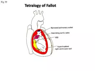
Tetralogy of fallot. First anatomic description… Danish anatomist Niels Stensen, in 1672... Described in detail by fallot in 1888…’la maladie bleue ’ Tetralogy of fallot with pulmonary atresia…10 %

E N D
First anatomic description…Danish anatomist Niels Stensen, in 1672... • Described in detail by fallot in 1888…’la maladiebleue’ • Tetralogy of fallot with pulmonary atresia…10% • Rarer variants include tetralogy of fallot with absent (or dysplastic) pulmonary valve and tetralogy of fallot with common atrioventricular canal[<5%]
Prevalence … 0.26 to 0.48 per 1,000 live births • In about 70% of tetralogy of Fallot patients, a putative genetic etiology remains to be determined. • Genes identified : NKX2.5, [4%]; JAG1 in Alagille syndrome.; TBX5 in Holt-Oramsyndrome. • Sibling recurrence rate …2.5% to 3% if only one sibling is affected…likely to increase substantially if more than one sibling is affected.
Environmental factors • maternal diabetes [threefold increased risk], retinoic acids, maternal phenylketonuria (PKU), and trimethadione Genetic cause : heterogeneous
Syndromes and associations….. DiGeorge/Velocardiofacial syndrome, Down syndrome, Alagille syndrome, cat's-eye syndrome, recombinant chromosome (or San Luis Valley) and Kabuki syndromes, and CHARGE and VATER/VACTERL associations. Microdeletionon 22q11….seen in • 15–35% of TOF patients; • 45% of TOF + PA; • 65% of those with TOF + APV.
Developmental FAULT • Defective embryonic neural crest migration abnormal conotruncal development. • Incomplete rotation and faulty partitioning of the conotruncus during septation…. • Malrotationof truncal-bulbar ridges results in misalignment of the outlet and trabecular septum and consequent straddling of the aorta over the malaligned VSD. • Abnormally anterior septation of the conotruncus by the bulbotruncalridgessubpulmonicobstruction
Tetrad …right ventricular outflow obstruction, aortic override, ventricular septal defect, and right ventricular hypertrophy PS Most characteristic …. subpulmonic stenosis • Obstruction along the entire course of the RVOT and pulmonary arteries (LPA commonly) can occur. • In general, the more severe the proximal obstruction, the greater the likelihood of distal areas of obstruction • Obstruction within the RV body : 1)hypertrophy of the septoparietal muscle bundles ; 2)anatomic displacement of the normal moderator band attachment
Pulm valve….bicuspid in 40% VSD : nonrestrictive…..Few ptsVSD restrictive d/t the accessory TV prolapsing thru the defect Coronary Anomalies • LAD from RCA (5%) & coursing in front of infund (Sx imp) • Single coronary (4%) So coronary evaluation before Sx imp • Echo….proximal coronary • If needed … root angio / CAG….. MRI/CT
LAD Pulm. Valve RCA AO RCC MPA PA NCC LCX LCx LAA RCA LAD
Right Aortic A in 25%......Twice more common in TOF+PA • Aortopulmonary Collateral Arteries..MC with TOF + PA…Only type 1, or bronchial artery collaterals, were documented in TOF with PS.: • Stenosis of LPA in 40% Associated cardiac abnormalities • PFO or a true atrial defect in 83% of hearts with TOF… AVSD (downs)…LSVC(11%) • Left heart lesions are rare…Aortic valve disease may be acquired as a result of surgical trauma, progressive root dilation, or 20to endocarditis. • TOF with Absence of PA…almost always LPA….PS murmur radiates to Rt chest…CXR..Lthemithoraxsmall,Lt lung hypovasc,Lthemidiaphragm elevated
Morphology of Pulmonary Arterial Supply • 3 major patterns : (1)most favourable… R/L PA are confluent, and are supplied by an arterial duct [unifocal] (2)the intrapericardialpulmonary arteries are confluent, but co-exist with systemic to pulmonary CAs. (3)absence of the intrapericardial pulmonary arteries….so fully supplied by multiple systemic-to-pulmonary CAs.
Unifocal….. usually the persistently patent arterial duct; rarely solitary systemic-to-pulmonary collateral artery/AP window/fistula from the coronary arteries. • Multifocal:MAPCAs: typically 2-6 in number; usually arise from the anterior wall of the aorta opposite the origin of the intercostal arteries. It is a rule that an arterial duct will not be present when a lung is supplied by systemic to pulmonary CAs. • The key to complete clinical diagnosis is to establish the course of each artery, to establish whether it runs directly into the lung or makes connections with intrapericardial and central pulmonary arteries, and to identify with precision the sites of these anastomoses. • Acquired CAs[TOF with PA] …. join the pulmonary circulation at immediately precapillarylevel.
APCs …the Rtupper artery supplies exclusively the RUL in direct fashion…Rtlower artery feeds the middle and lower lobes of the Rtlung through an anastomosis with the intrapericardialarterial tree, shown in blue, at hilarlevel….Left side…CAs are shown feeding the Lt lung through anastomoses at segmental level.
(1)Absent intrapericardial PAs (2)Anastomoses between the CAs and the intrapericardial PAs (extrapulm,hilar,lobar,segmental)
Pathophysiology and Hemodynamics • Severe cyanosis in profound RL shunting, • Some pts have a net LRshunt. • Hemodynamic features…RV hypertension because of the large VSD, with normal or low PAP.Thelow distal PAP is maintained as a result of the various levels of pulmonic obstruction. The PVR in the distal pulmonary arterial bed is usually normal. • The extent and direction of shunting ….determined by the cumulative amount of obstruction to PBF(Subpulmonic obstruction in all; obstruction @ valvular, supravalvular, and branch arteries are also common).
Balance between PBF & aortic BF will be determined by the difference in impedance between the unobstructed ,high-resistance systemic vascular bed and the obstructed pulmonary outflow tract and vascular bed. • RVH in proportion to LV mass in nonrestrictive VSD. • In restrictive VSD(suprasyst RVP)..severe RVH • Obstruction to PBF is relatively fixed & variation in cyanosis is determined by the changing SVR • Hypercyanotic episode…thought to result from an acute increase in infundibular obstruction and a proportional decrease in PBF.
C/F : • Wide due to variable severity of RV outflow obstruction • USGfetal diagnosis • Newborn & infants…cyanosis; systolic murmur • AcyanoticTOFmildpulmonary overcirculation rarely CHF unless there is a large PDA or aortopulmonary collateral arteries • Pregnancy…poorly tolerated … gest decrease in SVR increases R-L shunt & the labile SVR during labor,delivery leads to abrupt hypoxemia…..high fetal wastage / dysmature offspring
HYPERCYANOTIC SPELLS OR TETRALOGY SPELLS : • best described in TOF;canoccur with other forms of structural heart disease…. mediated, in part, by dynamic changes (acute increase)in subpulmonic obstruction….. changes in contractility due to ‘endogenous catecholamines or exacerbated by hypovolemia’ • other mechanisms[pulmonary atresia and VSD] …. decrease in systemic vascular resistance. • Child may assume squatting posture (instinctive) during spells … Pathogenetic mechanisms : Vulnerable respiratory control centres ;Increase in HR ; Increase in CO & VR ;Increase in R L shunt; Infundibular contraction may reinforce,but does not initiate
TYPICAL SPELL : • The child becomes distressed and inconsolable, without apparent reason, most often in the morning. • Peak incidence2nd-6th m ;few after 2 yrs • Crying is associated with progressively deeper cyanosis and hyperpnea (not tachypnea). • Spells are self-aggravating; • During the spell diminished/absent murmur • Not infrequently,the spell terminates with unconsciousness and, rarely,convulsions.If the hypoxemia is extreme, permanent neurologic sequelae and even death may ensue. True hypercyanotic spells are rare in neonates, although cyanosis may increase with crying.
O/E : • cyanosis ; clubbing ; • Arterial pulses …normal in uncomplicated TOF; • Wide pulse pressure (arterial diastolic runoff)…. aortopulmonary collaterals, palliative surgical shunt or PDA; • Accentuated precordial RV impulse; • LV impulse will not be hyperactive (normal cardiac output); • S2 single& loud (anterior, dextroposedaorta); • S3/S4 are unusual; • Aortic ejection click; • Systolic murmur…crescendo-decrescendo @ LUSB. The intensity of the murmur inversely parallels the degree of pulmonic obstruction; • Diastolic murmurs are unusual..Rarely AR murmur; • TOF with PA…..no harsh, obstructive precordial murmurs; • A harsh diastolic murmur, with a harsh murmur of PS, [harsh sawing, to-and-fro murmur ] ……TOF and APV syndrome; • Continuous murmurs….PDA;aortopulmonarycollaterals ;may be best heard in the back.
ECG • QRS axis … same as that of a normal newborn • RVH…Tall monophasic R in V1 with an abrupt change to an rS pattern in V2 (Tall R extends into adj precordial leads in TOF + APV) • Reduced PBF+ underfilledLVrS in V2-V6 • Balanced shunt …qR in V5,V6 • L-R shuntQR in V5,V6 • LAD with counterclockwise depolarisnTOF+AVSD
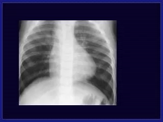
CXR: • Normal sized heart; [may be large in PA] • upturned apex; attenuated & concave left heart border (infundibularand PA hypoplasia)….boot-shaped heart, or coeur en sabot…small underfilled LV that lies above horiz IVS, inferior to which is a concentric hypertrophied nondilated RV • Diminished pulmonary vascularity in proportion to the degree of cyanosis. • Absent thymicshadow in the newborn may indicate associated chromosome 22q11.2 microdeletion(DiGeorge syndrome). • RAA in roughly 25%...accompanied by Rt DA on Rt side • In PA..lacy reticular pattern (d/t the anast b/w lobar/segm PAs & CAs) • Syst arterial collaterals rarely cause rib notching as they do not run in intercostal grooves.
ECHO: PLAX: • VSD; degree of aortic override; • ?bilateral conus; • AR; • Doppler… low velocity R-L shunt; • dilated CS…..?LSVC; • Absent DA..R/O RAA Cranial tilt of the transducer…..infundibulum and the proximal pulmonary arteries. [location and degree of infundibular, valvular, and arterial hypoplasia] Basal SAX: • RVOT and proximal pulmonary arteries…degree of PS….PDA? • VSD [just below the RCC, or at 10 o'clock position]. Tricuspid-aortic continuity confirms the perimembranousnature. • Coronaries…origin and course … echo showed sensitivity of 82%, specificity of 99%, and accuracy of 98.5% in a study by Need et al high parasternal and suprasternal views will provide visualization of the pulmonary arteries and aorta, respectively
LAD Pulm. Valve RCA RCC MPA NCC LCx The anomalous LAD crossing the RVOT in TOF is identified when the transducer is swept superiorly in the parasternal short -axis view . This allows visualization of the anomalous LAD that is situated anterior to the RVOT
Pulm valve Ao RCA Ao LAA Here a normal LAD (arising from the LCA and coursing posterior to the pulmonary valve) and a normal RCA are shown in the top frames for comparison.
64-slice HRCT : Questions about preoperative pulmonary artery anatomy, coronary artery anatomy, and systemic or pulmonary venous anatomy can frequently be resolved even in newborns with 64-slice high-resolution CT scans. CMR: Assessment of pulmonary anatomy Postoperative assessment of RV function, myocardial scarring, and PR fraction
Cardiac Catheterization • Less often needed ; • Diagnostic • Therapeutic • foremost goal …. clarification or better definition of anatomic characteristics, such as pulmonary arterial or coronary arterial anatomy • coronary artery anatomy … either by aortic root angio, selective coronary artery injection, or a combination of both. • Definition of any aortopulmonary collaterals ….usually originate from the descending aorta.
Medical Management • ductal-dependent pulmonary blood flow ?......neonates with critically restricted antegrade blood flow need to be started on prostaglandin E1and considered for either total repair or a systemic-to-pulmonary shunt. • Most newborns with TOF do not have ductal-dependent pulmonary blood flow and may be followed without specific early intervention. • Parental education for recognition of cyanotic spells [even acyanoticTOF are at risk for the development of spells in the first months of life]. • IE Prophylaxis for cyanotic/palliated patients ; patients for 6 months after patch repair surgery, and those with prosthetic valves
Hypercyanoticspells …Rx aims @ lowering impedance to pulmonary flow and further increasing systemic vascular resistance. Rx…..oxygen, volume expansion, sedation with morphine or ketamine, and, if needed, vasopressors[phenylephrine] Refractory to above ….. • Transfusion of whole blood/red cells • Balloon angioplasty of pulm annulus • Emergent surgical palliation or repair Propranolol …having some efficacy in minimizing or extinguishing the occurrence of spells. It may be used for patients who are awaiting surgical intervention or who have a medical contraindication to either complete repair or aortopulmonary shunt.
Infants with very severe RVOT stenosis and those with TOF +PA,with SaO2<70%, should have surgery within a few weeks after birth. • Infants with moderately severe stenosis and marked cyanosis (SaO270–90%) should have corrective surgery by 2–4 months. • Corrective surgery should be performed in all other infants with tetralogy of Fallot by 6 months. Contraindications for repair in early infancyPalliation initially • LAD from RCA crossing infundibulum • Severely hypoplastic PAs • Pulmonary atresia
Palliative Procedures Classic BT shunt[1945]…SCAPA on side opposite AA Modified BT… esp in small infants <6 months…side to side anastom with interposition graft of PTFE or Gore-Tex b/w SCA & PA [on the same side of AA] Waterston shunt : Side-side anastomosis of RPA to AA Potts : Side - side anastomosis of LPA to DA Waterston/Potts shunts : complications • Excessive PBF HF [20%] & PHTN • Difficulty taking shunt down at time of correction • Distortion of Rt/Lt PA ; Right/Left PA aneurysm
BT shunt-advantages : (a)low incidence of problems from excess PBF (b)No pericardial adhesions as pericardium is not entered (c)Easy to close @ time of complete repair..ligating its distal part just proximal to anastom with PA (d)Less distortion of PAs CENTRAL SHUNT:connecting a short tubular graft of Teflon or GoreTex from the aorta to the MPA. advantages vs other shunts: • the size of the communication could be controlled by selecting a tube with a diameter appropriate for the patient; • branch PAs are not disturbed so that reconstruction is not required at the time of corrective surgery.
Interventional procedures: • In patients with severe annular hypoplasia….. palliation of significant cyanosis by balloon valvuloplasty or RVOT stent placement can be done… Improvement in antegrade flow is thought to simultaneously enhance pulmonary arterial growth by augmenting PBF Balloon angioplasty of the pulmonary valve annulus is preferable to a shunt procedure as it is • less traumatic, • it avoids a thoracotomy, • reduces the likelihood of distortion of the pulmonary arteries • Coil embolization of APCs ….Coiling of vessels that perfuse pulmonary segments already supplied by pulmonary arterial flow serves to reduce LV volume loading as well as to eliminate runoff into the pulmonary arterial bed during CPB
Surgery aims @ • relieving all possible sources of RVOTO; • If possible, pulmonary valve function is preserved by avoiding a transannularpatch • Closure of VSD (dacron patch) • To relieve RVOTOpulmonaryvalvotomy, the insertion of an outflow tract patch or a transannular patch are often required. • Surgery during early infancy, when the pulmonary annulus is markedly stenotic, frequently requires the insertion of a long and wide transannular patch. • Consequently, most patients acquire PR as a result of the repair. PR may be well tolerated by many in the early postoperative years, but in the long term chronic PR is associated with reduced exercise capacity, RV dilatation, ventricular arrhythmias, and sudden death.
PVR : Early PVR in selected patients results in beneficial remodelling of the right ventricle Optimal timing is critical for preserving RV function (not too late) and avoiding the need for early re-operation (not too early). Amelioration of RV function following PVR has to be weighed against the risk of subsequent re-operation for homograft failure. Studies…RV end-diastolic volume may become a helpful indicator for defining both a lower limit (150 mL/m2) and an upper limit for re-intervention (200 mL/m2).[normal 60-100] CMR is the gold standard for evaluation of RV volumes and quantification of the degree of PR & TR.
SURGERY in TOF contd… VSD closure : Done thru’ RA approachwhether or not a trans-annular patch is used, as this approach allows to minimise the length of the right ventriculotomy (lengthonly necessary to relieve the RVOT obstruction and not for theVSD exposure). Efficacy of the RA approach …. • Preservesthe right ventricular function, ….. • Resultant PRafter limited transannular patching is less severe than thatwhich occurs after transventricular repair • Less incidenceof ventricular/atrial arrhythmias is • Easierto preserve the integrity & function of the tricuspid valve. Concerns of right ventriculotomy (classical RVapproach) • Low cardiac output in the early postoperativeperiod, • a higher incidence of arrhythmias, • Risk of late sudden death
In case of a major coronary artery crossing the RVOT…an external conduit or homograft would be necessary; Risk factors for early death after repair:1) very young age2) older age3) severity of annular hypoplasia4) small size of pulmonary arteries5) need for transannular patch (debatable)6) high peak RV to LV pressure ratio7) previous palliative operations8) multiple VSDs9) co-existing cardiac anomalies
Advantages of early total correction • Prevents cerebral hypoxia,cerebralembolism, abscess and hematological changes • Decrease RVPprevent persistent myocardial hypertrophy and probably reduce the risk of fibrosis of the RV • Providing adequate PBF will optimize the opportunity for normal growth of the main and branch PAs…also normal pulmonary circulation may be important for lung development. • Tendency for progressive hypertrophy of the RV infundibularregion is largely abolished by early repair. • Early separation of MAPCAs from aorta reduces the risk of PVOD
SURGERY IN TOF + PA : • PA anatomy not favourable? ….Palliative procedures… Central shunt vs reconstruction of RVOT using a patch/conduit while leaving open the VSD • If PA anatomy appears amenable to reconstruction, procedures leading to complete repair are indicated. Such procedures include RV outflow reconstruction for inducement of central pulmonary artery growth using a valved conduit / aortic or pulmonary homograft • If there is a connection between RV & PT correction can be achieved with a patch reconstruction
PA anatomy assessment • McGoonratio: (Diameter of RPA/DAo + Diameter of LPA/DAo) • Normal 2.1 • Adequate for VSD closure 1.2 • Inadequate <0.8 for VSD closure 2) Nakata Index:(CSA of RPA + CSA of LPA)/BSA • Normal value > 200 mm2/m2> 150 mm2/m2 is adequate.(Not usable preoperatively when MAPCAs are the major source of PBF & one-stage unifocalization + full repair is planned).
3) Total Neo-Pulmonary Artery Index (TNPAI) = APC index + Nakata Index • APC index is the sum of CSA of all usable APCs/BSA>250 - suitable for one-stage repair including VSD closure (These pts. have low RV/LV pressure ratio postoperatively). Critique of all these indices: These indices consider only the size of proximal vessel and not consider the condition of distal parts of the vessels (which may be stenosed). Recently the value of all these has been questioned
MAPCAs : management : 2 options 1)Obliterate them by Sx ligation/coil embolisation 2)Surgical unifocalisation : if it is the sole supply to many segments …..connecting all MAPCAs, as well as the small native pulmonary arteries, to one source of blood flow from the RV [ie to a central PA confluence or prosthetic PA confluence]….. If performed early,it avoids the dvpt of PVOD changes, as well as stenoses in the MAPCAs. If the pulmonary vascular morphology and resistance are such that adequate PBF can be accommodated…then RVP after correction will be low enough to close the VSD.
To assess whether this is likely, surgeons at the University of California in San Francisco perfuse blood from the perfusion system through the pulmonary artery at a rate of 2.5 L/min per m. If PA mean pressure is <25 mmHg, it is considered safe to close the VSD. If it is not closed, the patient is followed; the PVR may decrease over time, allowing later closure. Also, if stenoses in pulmonary vessels are noted, relief by balloon angioplasty, with stenting if necessary, may permit RVP to fall and permit closure of the defect. • Post surgery if the RVP/LVP is >0.75….RVF may result…avoid closure VSD / fenestrated patch closure
HypoplasticPAs … intervene early …. Encourage them to grow • Reconstruction of RVOT with a patch or valveless conduit • Placement of a central AP shunt • If MPA,RPA & LPA are present, even though very small (diameter 3 mm), they are capable of considerable enlargement if blood flow through them is increased. Creating an AP window early in infancy sufficient enlargement of the pulmonary arterial tree to later perform successful repair using the normal pulmonary arteries and a unifocalizationprocedure can be avoided.
Dvptof small pulmonary arteries (A) Before central shunt was performed, the Nakata index was 64 and the McGoon ratio was 0.82. (B) One hundred seventy-four days after operation, the Nakata index rose to 89 and the McGoon ratio to 1.1.
Post Repair for TOF in general • Current surgical survival, even for symptomatic infants <3 months of age, is excellent. • Hospital and 1-month survival rates of 100% have been reported. • Earlier age at repair (<1 year of age) did not adversely affect the rate of reintervention; so primary repair should be regarded as the preferred management strategy. • Twenty-year survival for hospital survivors, irrespective of management strategy, was 98% for patients who have TOF with PS and slightly lower for patients with PA, reflecting the overall excellent long-term survival of these patients.