Download
1 / 17
390 likes | 920 Views
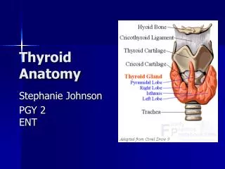
THYROID ANATOMY. Nani M Yazid, drg,.M.Kes Departement Of Anatomy. Background. What: brownish-red, highly vascular gland Location: ant neck at C5-T1, overlays 2 nd – 4 th tracheal rings width: 12-15 mm (each lobe) height: 50-60 mm long

E N D
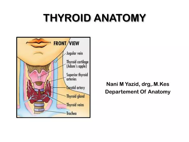
THYROID ANATOMY Nani M Yazid, drg,.M.Kes Departement Of Anatomy
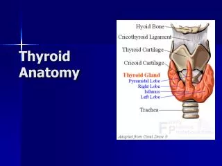
Background • What: brownish-red, highly vascular gland • Location: ant neck at C5-T1, overlays 2nd – 4th tracheal rings • width: 12-15 mm (each lobe) • height: 50-60 mm long • weight: 25-30 g in adults (slightly more in women) **enlarges during menstruation and pregnancy**
Relation Strap muscles • Lateral - sternothyroid • Anterior • omohyoid muscle • sternohyoid • Inferior - SCM (lower portion)
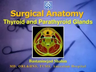
THYROID GLAND • Location: Located close to thyroid cartilage. Has two lateral lobes connected by thyroid isthmus medially. Isthmus covers cricoid cartilage in ventral view. • Development: first endocrine gland to apear during development. Develops from endodermal thickening in floor of early pharynx and epithelium of 3rd and 4th gill slit pouches as early as 24 days after fertilization. Starts out caudal to tongue, but ultimately comes to be wrapped around laryngeal cartilages.
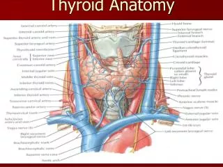
THYROID GLAND • Arterial Supply: superior thyroid artery (branch of external carotid artery). • Venous Drainage: drained by dense interconnected network of pharyngeal veins that eventually dump into superior thyroid vein and inferior thyroid vein. These are tributaries of intrenal jugular veins and left brachiocephalic vein respectively. • Functions: • THYROXIN –regulate rate of metabolism • CALCITONIN –decreases levels of calcium and phosphate in the blood (partially antagonistic to parathyroid hormone).
VENOUS: 3 pairs of veins: • STV – asc along STA and becomes a tributary of the IJV • MTV – directly lateral IJV • ITV Lymphatics
Lymphatics • Extensive, multidirectional flow • periglandular prelaryngealpretracheal paratracheal (along RLN) brachiocephalic (sup mediastinum) deep cervical thoracic duct
Innervation Principally from ANS • Parasympathetic fibers – from vagus • Sympathetic fibers – from superior, middle, and inferior ganglia of the sympathetic trunk Enter the gland along with the blood vessels.
PARATHYROID GLAND • Location: • Usually paired. • Very small (less than 5 mm). • Called parathyroid glands because of their position on posterior margins outer surface of thyroid gland. • More superior of each pair usually near middle of margin of lobe. • More inferior of each pair usually at inferior apex of lobe. • Development: Like thyroid gland, develop from endodermal thickening in floor of early pharynx and epithelium of 3rd and 4th gill slit pouches
Innervation, Vascularization: same as thyroid gland. • Function: • PARATHYROID HORMONE (PTH) – raises the level of calcium in the blood, decreases levels of blood phosphate. Partially antagonistic to calcitonin of thyroid gland.