Download
1 / 46
460 likes | 636 Views
RADIATION PROTECTION TRAINING. Charles F. Reindl, M.S., C.H.P. Certified Diagnostic Radiological Physicist Radiation Safety Officer Tulane University Office of Environmental Health & Safety. Section 1 Radiological Fundamentals: Matter Radioactive Decay & Types of Ionizing Radiation

E N D
RADIATION PROTECTIONTRAINING Charles F. Reindl, M.S., C.H.P. Certified Diagnostic Radiological Physicist Radiation Safety Officer Tulane University Office of Environmental Health & Safety
Section 1 Radiological Fundamentals: Matter Radioactive Decay & Types of Ionizing Radiation Radiation Interactions Radiation Exposure Units External Exposure Measurement Biological Effects of Radiation Federal Limits for Occupational Exposure to Ionizing Radiation Section 2 Instrumentation & Radiation/Contamination Monitoring: Gas-Filled Detectors Radiation Monitoring Contamination Monitoring Survey Frequency Other Laboratory Rules Section 3 Exposure Reduction: Inverse Square Law Time Shielding Section 4 Radioactive Decay & Specific Hazards: Decay Equation Radioiodine Tritium Phosphorus-32 Section 5 Radioactive Materials Disposals: Decay to Background Levels Sewer Disposal Incineration Transfer to a Licensed Disposal Firm Table of Contents:
Radiological Fundamentals Section 1
Section 1 Radiological Fundamentals • Matter • Radioactive Decay & Types of Ionizing Radiation • Radiation Interactions • Radiation Exposure Units • External Exposure Measurement • Biological Effects of Radiation • Federal Limits for Occupational Exposure to Ionizing Radiation
Section 1-AMatter A. Matter • All matter is composed of atoms, and each atom is made up of three fundamental particles. SymbolNameMassCharge p+ Proton 1 amu +1 e- Electron 0.0005 amu -1 n Neutron 1 amu 0
An amu (Atomic Mass Unit) is approximately equal to the mass of a proton or neutron and numerically equal to 1.66 E-24 grams. The mass of an electron is negligible in comparison. Any atom can be symbolized by the following notation: AXZ where: X = The chemical symbol of the element which is determined by the number of protons in its nucleus. Z = The Atomic Number, equal to the number of protons in its nucleus. A = The Mass Number, equal to the number of protons plus neutrons in its nucleus. Section 1-A, continuesMatter
Section 1-A, continues Matter • There are three isotopes of the element hydrogen, symbolized "H.“ • The first isotope is H-1, the most common type of hydrogen with a natural abundance of 99.985%. • The second isotope is H-2, heavy hydrogen, also called deuterium with a natural abundance of 0.015%. • The final isotope is H-3, radioactive hydrogen, also called tritium. • All three are isotopes of hydrogen and H-3, since it is radioactive, is also called a radioisotope or a radionuclide. In general, isotopes are atoms with the same number of protons (same Z), but different numbers of neutrons (different A).
Section 1-BRadioactive Decay & Types of Ionizing Radiation B. Radioactive Decay and Types of Ionizing Radiation Radioactive atoms become more stable by emitting one or more of the following most common types of ionizing radiation: • Alpha (α): A high speed helium nucleus (4He2++) with no orbital electrons and a resulting +2 charge overall. • Beta (β): A high speed electron (e-) with a -1 charge overall. • Gamma (γ): An electromagnetic wave with no mass and no charge overall.
Section 1-CRadiation Interactions C. Radiation Interactions • All ionizing radiations produce ion pairs as they travel through air, detection devices, shielding, or body tissue. These ion pairs are simply target atoms whose electrons have been stripped off by the ionizing radiation. If Y is any target atom: Y + ionizing radiation -----> Y+ + e- where the two products are the ion pair. The formation of ion pairs may result in ionization of the air, ionization causing a pulse in a detector, heating of shielding, or biological damage depending on what the target atom is. • Particles with electric charge such as alphas and betas pull or push target electrons through charge-charge interactions (unlike charges attract while like charges repel) as they lose energy slowing down. • Ionizing electromagnetic waves such as gammas or X-rays (X-rays being simply low energy gammas) produce ion pairs by photointeractions producing recoil betas/electrons which go on to produce further ion pairs by charge-charge interactions.
Section 1-DRadiation Exposure Units D. Radiation Exposure Units Radiation exposure can be measured by use of the following units, listed in order from oldest to most modern: • Roentgen (R) - The amount of gamma or X-radiation producing one esu (electrostatic unit) of ion pairs in a cubic centimeter (cc) of dry air. This amounts to about two billion ion pairs in a cc of air. • This same amount of gamma or X-radiation produces approximately one. • Rad (Rad) - The amount of any type of radiation depositing 100 ergs of energy per gram of any material. This amounts to about two trillion ion pairs in a gram (g) of air, one gram of air being about a thousand cubic centimeters, and about the same number of ion pairs in a gram of body tissue.
Section 1-D, continuesRadiation Exposure Units • Numerically, one Rad = 100 ergs/g of absorbed radiation. • One Roentgen of gamma or X-radiation produces about one Rad (100 ergs/g) of absorbed dose and also one. • Rem (rem) - The amount of any type of radiation producing biological damage equivalent to that deposited by 100 ergs of gamma or X-radiation per gram of body tissue. In other words, a rem of any type of radiation will always produce the same amount of harm to living tissue as would a Rad of gamma or X-radiation. The damage produced by gamma or X-radiation becomes the standard by which all other types of radiation are measured. • Numerically, one rem = the dose equivalent to 100 erg/g of or X-rays.
Section 1-D, continuesRadiation Exposure Units • Gammas and x-rays generally cause a spray of recoil electrons (betas) when they interact with cells through photointeractions. These recoil betas, because of their -1 charge and high speed, then produce relatively dispersed damage among many cells. Because (γ/Χ-radiation produces recoil betas, β/γ/Χ-radiation produce the same number of Rads and rems. • The Roentgen, Rad, and rem are relatively large units relative to research laboratory work, so subunits in the milli (one thousandth) range are frequently employed. Note that: 1 R = 1000 mR 1 Rad = 1000 mRad 1 rem = 1000 mrem
Section 1-EExternal Exposure Measurement E. External Exposure Measurement • Alphas will never penetrate the outer layer of dead skin if present in an external radiation field and are therefore never counted as external exposure. However, ingested or inhaled alpha sources will deliver their entire absorbed radiation dose. • Betas will penetrate as far as living skin if present in an external radiation field, contributing to external skin/shallow dose. • Gammas, having no charge to interfere with their progress, will penetrate living skin, internal organs, and possibly re-emerge from the far side of the body because of their great range. They are counted as part of the skin/shallow and whole body/deep dose because their recoil betas can be produced anywhere and impart biological damage at that location. Note:An "M" reading under deep/whole body or shallow/skin exposure reported on a Landauer Radiation Dosimetry Report means "Less than the Minimum Detectable Exposure."
Section 1-FBiological Effects of Radiation F. Biological Effects of Radiation • Cell sensitivity to radiation is determined by two primary factors: • Level of cell activity resulting in increased rates of chemical diffusion across the nuclear membrane. • Rate of cell division resulting in more time spent with the protective nuclear membrane dissolved. • Other factors apply but will be discussed later.
Section 1-F, ContinuesBiological Effects of Radiation • These criteria can be used to list the body's cells and organs in approximate order from most to least radiosensitive: • Fetal tissue • Reproductive cells (for long term genetic reasons). • Red and white blood forming cells primarily located in the bone marrow. • Lens of eye. • Most internal organs such as the lung and lower intestine. • Skin of the whole body, thyroid, nerve, etc. • Extremities such as hands and feet. • In order to put the rem into its proper biological perspective, it is useful to compare it to the effects of large acute exposures which are received in 24 hours or less.
Section 1-F, ContinuesBiological Effects of Radiation • CAUTION: The exact boundary of each exposure range depends on individual health and the availability of medical treatment after exposure. rem Immediate Effects 0 - 25 None 25 - 100 Small measurable changes in white blood cell count. 100 - 200 Possible symptoms of radiation sickness: Blood changes including a white blood cell decrease leading to decreased disease resistance, a red blood cell decrease leading to fatigue, and a blood platelet decrease leading to a decreased ability of blood to clot over wounds. Intestinal wall damage leading to nausea, vomiting, and diarrhea.
Section 1-F, ContinuesBiological Effects of Radiation • Note: The severity of symptoms increases with increasing exposure until the following approximate exposures are reached: 500 Lethal Dose to 50% if those exposed within 30 days (LD50/30) along with epilation (loss of hair) within two weeks. 1000 Additional symptoms include convulsions due to Central Nervous System damage. • The American Cancer Society states that 25% of the 20 to 65 year old age group develops cancer from sources such as errors in gene duplication, smoking, air pollution, food, and natural background radiation. An increased exposure of 1 rem would increase the risk of cancer from about 25% to about 25.03%.
Section 1-F, ContinuesBiological Effects of Radiation • This assumed linear relationship between radiation exposure and the risk of effects provides the rationale for Federal limits on radiation exposure to the whole body including bone marrow, genetic material, trunk and head. • A "background" exposure rate exists naturally. In the U.S., the natural background radiation exposure rate is about 100 mrem/year from traces of naturally occurring Uranium and Thorium in soil and building materials, traces of naturally occurring K-40 and C-14 in foods, and cosmic radiation. Including an average number of medical and dental X-rays, the total U.S. average increases to about 200 mrem/year.
Section 1-GFederal Limits for Occupational Exposure to Ionizing Radiation G. Federal Limits for Occupational Exposure to Ionizing Radiation • The occupational limits set forth in Title 10, Code of Federal Regulations, Part 20 (10 CFR 20) and Louisiana State Regulations apply to those with a complete prior occupational radiation exposure history. • The limit for occupational exposure to ionizing radiation is 5,000 mrem/year. • The Federal limit for pregnant women is based on exposure to the fetus which is very radiosensitive. • The limit for radiation exposure to the fetus is 500 mrem/term.
Instrumentation andRadiation/Contamination Monitoring Section 2
Section 2 Instrumentation and Radiation/Contamination Monitoring • Gas-Filled Detectors • Radiation Monitoring • Contamination Monitoring • Survey Frequency • Other Laboratory Rules
Section 2-AGas-Filled Detectors A. Gas-Filled Detectors • Once again, radiation interactions produce ion pairs. Gas-filled detectors generally consist of an outer container along with an inner wire placed inside and along the long axis of the detector probe. Container (outer electrode) and wire (inner electrode) are electrically insulated while the outer container is generally given a negative charge and the inner wire a positive charge. • With voltage on the electrodes, an electric field is created which attracts the negatively charged electrons to the positive inner electrode and the positively charged ions to the negative outer electrode. A voltage drop or current increase is produced in the attached detector circuitry which is amplified and counted as a pulse.
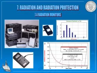
Section 2-BRadiation Monitoring B. Radiation Monitoring Radiation levels must be measured in order to determine the rate at which dose is being received. This can only be done by using radiation survey meters. Before performing a radiation survey, the following preoperational checks of the survey meter are recommended: • Battery check the instrument by turning the range selection switch to the battery check position to see if the meter measures adequate voltage in the "Battery O.K." region. • Source check the instrument with a check source on contact with the meter probe. The meter should respond to radiation.
Section 2-CContamination Monitoring C. Contamination Monitoring • Activity (amount of radioactive material) can be measured in units or subunits of the Curie (Ci). Note that: 1 Ci = 1,000 mCi = 1,000,000 μCi Example: An incoming source vial contains 0.25 mCi of P-32. How many μCi is this? 0.25 mCi X 1,000 μCi/mCi = 250 μCi • Loose contamination can be picked up and spread to other areas. It is measured by taking a "wipe" such as a piece of filter paper, cloth, or a cotton-tipped swab and rubbing it over a specified area. Most frequently a 100 cm2 area which is about 4 inches by 4 inches is used as a standard. The smear is then beta or gamma counted. The loose activity is then calculated as in the following example:
Section 2-C, continuesContamination Monitoring • Example: A cotton-tipped swab wiped over 100 cm2 area of a fume hood surface produces 50,000 counts per minute (cpm) above background on contact with a survey meter. The Conversion Factor on the calibration sticker of the meter reads "1 μCi = 100,000 cpm". What is the loose surface contamination level in the fume hood? 50,000 cpm X 1 μCi/100,000 cpm = 0.5 μCi/100 cm2 • A laboratory sample counter can be used to measure lower levels of contamination more accurately, as long as a standard with a known number of μCi is used to determine the conversion factor from μCi to cpm. This is the same as determining the efficiency of the counter.
Section 2-DSurvey Frequency D. Survey Frequency • Laboratories which use radioactive materials continuously must be surveyed weekly and the survey results recorded. Infrequent use laboratories must be surveyed at the completion of the procedure. • A record of these surveys, even if negative, must be kept on file for inspection by State or Federal regulators.
Section 2-EOther Laboratory Rules E. Other Laboratory Rules • Decontamination of work areas must be performed when contamination levels exceed twice background. • Refrigerator/freezers that are used to store radioactive materials must be labeled “Caution, Radioactive Materials.” • Incoming radioactive material packages labeled White I, Yellow II, or Yellow III must be wipe tested for radioactive contamination and the results recorded. This requirement is not applicable if the packages arrive at 333 South Liberty Street since the Radiation Safety Office checks these packages for contamination on a daily basis. • Radioactive materials must be secured when not in use by storing them in a locked container or locking the laboratory door when absent from the room.
Exposure Reduction Section 3
Section 3 Exposure Reduction • Inverse Square Law • Time • Shielding
Section 3-AInverse Square Law A. Inverse Square Law • The Inverse Square Law for gamma point sources is: D1 X r12 = D2 X r22 • Example: The dose rate one foot away from a point source is 100 mrem/hr. What is the dose rate after stepping back to a distance of two feet? D2 = (100 mrem/hr) X (1 ft)2/(2 ft)2 = 25 mrem/hr • As can be seen from the previous example, doubling distance from a point source of radiation decreases dose rate to one quarter of what it was. This is the basis of the Inverse Square Law and dose reduction by increasing distance.
Section 3-BTime B. Time • The time equation, which is applied frequently in radiation protection work, is: Dose = D x T where: D = Dose rate T = Time of exposure Example: A researcher stands in an area where a survey meter reads 50 mrem/hr for a period of six hours. What is their total exposure as a result? 50 mrem/hr X 6 hr = 300 mrem
Section 3-CShielding C. Shielding • The range of a beta is sufficient to penetrate living skin. Because of its -1 charge, a few millimeters of plastic can stop all betas. • Recall that gammas and X-rays are electromagnetic waves with no mass or charge and very penetrating. One Half Value Layer (HVL) is the thickness of shield material that will reduce exposure rate to one half of its initial amount. The thickness that reduces the incident flux to one half will, if doubled in thickness, reduce the original incident flux to one quarter of what it was. • In equation form: D = D0 (1/2)n and n = x/HVL where: D0 = Unshielded dose rate. D = Shielded dose rate. n = Number of Half Value Layers. x = Shield thickness.
Section 3-C, continuesShielding Example: A source is producing a dose rate of 100 mrem/hr at the side of a laboratory bench. Estimate the remaining dose rate from the source if two 1/2 inch lead shields are placed over the source. The HVL is 0.5 inch for the gamma energy involved. The total thickness of lead shielding is 1.0 inch and n = 1.0 in/0.5 in = 2 D = (100 mrem/hr) X (1/2)2 = 25 mrem/hr
Radioactive Decay and Specific Hazards Section 4
Section 4 Radioactive Decay & Specific Hazards • Decay Equation • Radioiodine • Tritium • Phosphorus-32
Section 4-ADecay Equation A. Decay Equation • The following is the decay equation: A = A0(1/2)n where: A0 = Activity initial. A = Activity final. n = t/t1/2 = Number of half-lives that have elapsed. t = Time that has elapsed. Example: A radioactive sample has a half-life of 30 minutes. If the sample initially contained 1 mCi, how much remains after 60 minutes? n = 60 min/30 min = 2 elapsed half lives A = 1 mCi X (1/2)2 = 0.25 mCi • The example is simple but illustrates the point that one half-life of time will decrease a given amount of radioactivity to one-half of what it was.
Section 4-BRadioiodine B. Radioiodine Radioiodine is most commonly I-125 with a 60 day half-life. It produces a relatively low energy 35 keV gamma/x-ray in only 7% of the decays. When purchased as sodium iodide (NaI) in base (NaOH) the radioiodine is relatively stable and water soluble. Under acidic conditions, sodium iodide chemically partitions to form volatile elemented iodine (I2) which can be inhaled. Due to its rapid biological accumulation in the thyroid, thyroid monitoring is necessary. Radioiodine is primarily an airborne thyroid hazard.
Section 4-CTritium C. Tritium • Tritium, H-3, has a 12.3 year half-life. • It produces a 19 keV beta and no gamma. • Tritium is an internal exposure hazard. • Rubber gloves must always be used when handling radionuclides. • Including tritium since it can be absorbed through bare skin. • Urinary monitoring, which is done by collecting a milliliter of urine to be mixed with scintillation medium and counted, must be performed when using large amounts of tritium.
Section 4-DPhosphorus-32 D. Phosphorus-32 • Phosphorus-32 (P-32) has a 14.3 day half-life and emits a 1,710 keV beta with no gamma. • P-32 is an external skin/shallow exposure hazard, while not a whole body/deep dose hazard. • It can also deliver a large dose to the hand when handled in mCi amounts.
Radioactive Materials Disposal Section 5
Section 5 Radioactive Materials Disposal • Decay to Background Levels • Sewer Disposal • Incineration • Transfer to a Licensed Disposal Firm
Section 5-ADecay to Background Levels A. Decay to Background Levels • Radioactive waste may be discarded in the normal waste stream if the following conditions are met: • The material has decayed 10 half-lives. • The waste produces no count rate above background on contact with a radiation survey meter. • All "Radioactive" labels have been removed/defaced. • Notation is made on the "Radionuclide Receipt & Use Record" that the waste has been defaced & discarded, ≤bkg, and date. Note: Accurate Radionuclide Receipt and Use Records prove to Federal and State auditors that radioactive material has been used and disposed of in a safe and legal manner. • After decay, the waste may still be hazardous or infectious and have to be disposed of in one of these specialized waste streams.
Section 5-BSewer Disposal B. Sewer Disposal • Aqueous liquid radioactive waste may be disposed of in the laboratory "hot sink" along with copious amounts of water as long as the date, radioisotope, and activity disposed of is recorded. • Its very important to note that this method may only be used if the sewer is connected to a municipal sewer system. • A "Semi-Annual Radionuclide Inventory/Sewer Disposal" report is requested of each licensee every six months. These are used by the Radiation Safety Office to ensure that the activity concentrations leaving each building are below drinking water concentration limits and that the total activity discharged is less than 1 Ci per year.
Section 5-CIncineration C. Incineration • Contaminated laboratory trash and biodegradable liquid scintillation vials can be taken to the Medical School waste room, tagged with the radioisotope, activity amount in µCi, date, and licensee name for eventual incineration. • The Waste Room is located in room 1105 of the Medical School and is open Tuesdays from 8:30 to 10:30 a.m. • The Radiation Safety Office ensures that the amount of radioactivity incinerated per hour does not cause effluent air concentrations to exceed the limit for unrestricted air and that the resulting incinerator ash does not exceed drinking water concentration limits.
Section 5-DTransfer to a Licensed Disposal Firm D. Transfer to a Licensed Disposal Firm • Organic liquid scintillation cocktail such as toluene containing radioisotopes with long half-lives must be transferred to a licensed radioactive/chemical waste disposal company. • The Radiation Safety Office will arrange these shipments, but must bill the licensee generating the waste because of the high price of such disposal.
RADIATION PROTECTION TRAINING Proceed to Quiz https://audubon.tulane.edu/ehs/enterssn.cfm?testnum=134