Download

1 / 47
470 likes | 945 Views
Novak 2002. Endometrial cancer. Introduction Epidemiology and risk factors Endometrial hyperplasia Screening for endometrial Ca. = Most common malignancy in the ♀ genital tract = ½ ♀ genital tract malignancy in USA

E N D
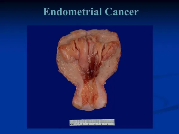
Novak 2002 Endometrial cancer
Introduction • Epidemiology and risk factors • Endometrial hyperplasia • Screening for endometrial Ca
= Most common malignancy in the ♀ genital tract = ½ ♀ genital tract malignancy in USA = 4th most common cause of malignancy after breast, lung, and bowel = 7th leading cause of death from M = 2 – 3 % in the female introduction
Recently ↑ awareness of EC due to: • ↓ Ca cervix • ↑ life expectancy • HRT • Earlier diagnosis due to: - Easier diagnostic tools - Understandingof premalignant lesions
EC occur primary in postmenopausal women and virulence ↑ by age • Although EC is usually presented in an earlier stage, deaths from EC > that from cervical cancer • Unopposed estrogen ↑ risk of EC
In the past decades management of EC evolved from: • Preoperative intrauterine radium packs • External pelvic irradiation followed 6 months later by hysterectomy • Single brachytherapysession followed by hysterectomy • Hysterectomy and PO ttt depending on surgical or pathological findings
EC is 2 types: (1) (2) Young Old Hormone-dependent Hormone-independent Due to hyperplasia Due to atrophy Well differentiated Undifferentiated Good prognosis Poor prognosis The 2nd type ↑ in: Elderly – thin – obese – postmenopausal Epidemiology and risk factors
Risk factors: • Unopposed estrogen as in: - Nullipara X 2 – 3 times - Infertile - Anovular - Irregular bleeding • Overweight 21 – 50 pounds X 3 “ > 50 pounds X 10 “
Menopause > 52 years X 2.4 times • PCO • Functioning ovarian tumors • HRT X 4 – 8 times ↑ risk by ↑ dose and duration • Tamoxifen X 2 - 3 • DM X 1.3 – 2.8 times • HTN and hypothyroidism
A spectrum of: biological/ morphological alteration in endometrial gland/stroma Ranging from: exaggerated physiological changes to cancer insitu Due to: estrogen over stimulation of the endometrium without progesterone Endometrial hyperplasia
EH may be associated with: • Bleeding • Functioning ovarian tumors • HRT • EC Recent classification depends on: • Architectural features • Cytological features
Architectural depends on: • Crowdening of the glands • Complexty of the glands Non atypic EH: • Simple: Cystic dilatation of the glands Slightly irregular glands No atypia
Complex: Crowedening, budding, infulding of the glands with less stroma Atypia: Nucleus = large, variable in size & shape Cytoplasm = ↑ nuclear/cytoplasmic R = loss of polarity Nucleolus = prominent
Chromatin = irregular, clumping = parachromatin clearance Transformation to malignancy: Nonatypic: Simple 1% Complex 3% Atypic: Simple 8% Complex 29%
ER is stable in 18% regress in 74% If D&C show atypia Hystrectomy show EC in 25% of cases Prognosis depends on: • Age • Ovarian disease • Exogenous hormones • Obesity • Endocrine function
MPA 10 – 20 mg/day treatment results: NonatypicAtypic Regression 84% 50% Recurrence 6% 25% Malignancy 25% Megestrol acetate treatment in atypia: Regression = 93% Recurrence = 20%
Progestines treatment of EH is less effective in atypic EH than nonatypic EH Treatment in nonatypic EH: • Ovulation induction • Cyclic progestines: MPA 10 – 20 mg/day for 14 days/month Treatment in atypic EH: • Continues progestines
Megestrol acetate: 20 – 40 mg/day X 2 – 3 months followed 3 – 4 weeks by EB Follow up by U/S or EB is important due to: • 25% undiagnosed malignancy • 29% malignancy transformation • ↑ recurrence rate
No screening test is: appropriate acceptable cost effective ↓ mortality • Pap smear = inadequate • Cytology = ↓ sensitivity & specificity • Progesterone challenge test = only Screening of endometrial Cancer
for estrogen primed endometrium • TVS & EB = too expensive Screening discover only 50% of EC Screening of high risk women: • HRT without progesterone • Familial nonpolypoid colon cancer Pap smear is +ve only in 30 – 50% of EC
90% of EC represent by abnormal bleeding • Some EC complain of pelvic discomfort or heaviness = uterine enlargement or extrauterine extension • Elderly women with cervical stenosis complain of excessive offensive discharge with no bleeding = hematometria or pyometria poor prognosis symptoms
5% of EC are asymptomatic and discovered accidently by: - Abnormal pap smear in advanced cases - CT/ US for other reasons - Hysterectomy for other reasons Any perimenopausal bleeding must be evaluated even if minimal or recurrent
Causes of vaginal bleeding: • Nongenital • Genital extrauterine • Uterine Extrauterine causes are evaluated by: • History • Examination • Search of blood in urine/stools
Vulval/vaginal/cervical lesions: Can be seen and biopsy is taken Vaginal atrophy = 15% of vaginal bleeding thin and friable vagina Causes of uterine bleeding: • Endometrial atrophy = 60 – 80% EB insufficient endometrial tissue
( blood and mucus only ) No bleeding after EB • Polyps: = 2 – 12% Difficult to diagnose by D&C or EB Diagnosed by hystroscopy, TVS or sonohystrography If not diagnosed unnecessary hystrectomy
HRT: = 15 – 25% X 4 – 8 ↑ risk of EC ↑ risk by ↑ duration and dose ↓ risk by progesterone and follow up by TVS and EB annually and if recurrent bleeding occur • Hyperplasia = 5 – 10% • Cancer = 10%
Bleeding in EC: • Menometrorrhagia • Oligomenorrhia • Cyclic > menopausal age Cancer is suspected if: • Persistent • Recurrent • Obese • anovular
General examination: Obesity – HTN Breast - Peripheral LN Abdominal examination in advanced EC: • Ascitis • Nodular liver • Nodular omentum signs
Genital examination: - Inspection & palpation: Vaginal introitus Vagina Suburetheral area Cervix - Bimanual rectovaginal examination: • Uterus size – mobility • Adnexa masses • Parametrium induration • Doglas pouch nodularity
I – Office endometrial aspiration: Accurate in 90 – 98% • Inexpensive • No tenaculum • No cramps • Well tolerated • Adequate tissue sample diagnosis
2 – Pap smear: unreliable +ve in 30 – 50% of EC 3 - Hystroscopy/D&C for: • cervical stenosis • patient intolerance • inadequate tissue sample • recurrent bleeding with –ve EB
4 – TVS/EB: To select patients for hystroscopy or sonohystrography Patents with endometrial thickness ≥ 5 mm or with polypoidal mass or fluid require further evaluation
Symptoms • Signs • Diagnosis • Pathology • Preoperative evaluation • Staging • Prognostic variables
90% 0f the patients represent by abnormal bleeding • Some women complain of heaviness and pelvic discomfort = uterine enlargement or extrauterine extension • Some elderly women may have cervical stenosis hematometria – pyometria – offensive discharge = poor prognosis symptoms
5% of patients are asymptomatic and discovered accidently at: - Pap smear advantage stage - Hystrectomy for other reason - CT/ US for other reason Any perimenopausal bleeding should be evaluated even if minimal or nonpersistant Causes of bleeding may be:
Nongenital • Genital but extrauterine • Uterine Nongenital causes are diagnosed by: C/P + C/E + blood in urine/stool Vaginal atrophy = 15% of vaginal bleeding Thin and friable vagina Uterine cause should be excluded
Causes of uterine bleeding: • Atrophy 60 – 80% • HRT 15 – 25% • Polyp 2 - 12% • Hyperplasia 5 - 10% • Cancer 10% Atrophy: usually occur in women > 10 years postmenopausal
EB insufficient tissue ( blood + mucus) no bleeding after EB Polyp: difficult to diagnose by EB/D&C Hystroscopy/sonohystrography better If not diagnosed unnecessary hystrectomy HRT: ↑ risk X 4 – 8 times if no progesterone ↓ risk by progesterone and follow up by annual TVS/ EB
Cancer: Present by abnormal bleeding: • Menometrorrhgia • Oligomenorrhia • Cyclic bleeding beyond menopause Consider malignancy if: • Persistent /recurrent bleeding • Obese/anovular patient
General examination: Obesity – HTN Breasts – peripheral LN Abdominal examination: Ascetis Nodular liver/omentum Genital examination: - Inspection & palpation: signs
Vaginal introitus Vagina Suburethral area Cervix - Bimanual rectovaginal examination: • Uterine size/mobility • Adenexal masses • Parametrialinduration • Doglas pouch nodularity
I – Office endometrial aspiration: Accurate in 90 – 98% • Inexpensive • No tenaculum • Minimal uterine cramps • Well tolerated • Adequate tissue sample in 90% diagnosis
- If cervical stenosis paracervical block cervical dilatation - If cervical pathology suspected endocervical sample - Premedication by antiprostaglandins II – Pap smear: Unreliable +ve in 30 – 50% of EC
III – Hystroscopy/ D&C: Indicated in: • Cervical stenosis • Patient intolerance • Inadequate sample • Recurrent bleeding with –ve EB IV – TVS / sonohystrography:
Helps to select patients with minimal or adequate endometrial thickness Evaluated any patient with: • Endometrial thickness > 4 mm • Polypoidal mass • Fluid