Download
1 / 20
200 likes | 318 Views
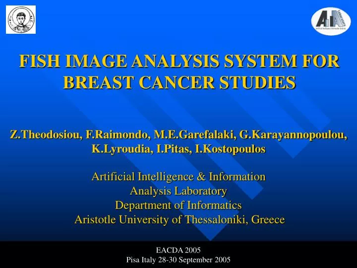
FISH IMAGE ANALYSIS SYSTEM FOR BREAST CANCER STUDIES. Z.Theodosiou, F.Raimondo, M.E.Garefalaki, G.Karayannopoulou, K.Lyroudia, I.Pitas, I.Kostopoulos . Artificial Intelligence & Information Analysis Laboratory Department of Informatics Aristotle University of Thessaloniki, Greece. EACDA 2005

E N D
FISH IMAGE ANALYSIS SYSTEM FOR BREAST CANCER STUDIES Z.Theodosiou, F.Raimondo, M.E.Garefalaki, G.Karayannopoulou, K.Lyroudia, I.Pitas, I.Kostopoulos Artificial Intelligence & Information Analysis LaboratoryDepartment of Informatics Aristotle University of Thessaloniki, Greece EACDA 2005 Pisa Italy 28-30 September 2005 AIIA Lab, Department of Informatics Aristotle University of Thessaloniki
Introduction • The evaluation of fluorescent in situ hybridization (FISH) images is a method used to determine Her-2/neu status of breast samples. • FISH technique allows the analysis and quantification of specific abnormalities (gene amplifications). • DNA probes target the HER-2/neu gene and attach themselves to their target sequence. This process is called hybridization. • The probes carry special fluorescent markers that emit light, when the probes bind to the HER-2 genes. AIIA Lab, Department of Informatics Aristotle University of Thessaloniki
Problem Statement • FISH images:The Her-2 probes are visible as orange stained spots. Probes for centromere 17 (CEP-17), are visible as green spots. The sections are counterstained with DAPI, providing a blue background for nucleus body. AIIA Lab, Department of Informatics Aristotle University of Thessaloniki
Problem Statement (2) • FISH images conventional analysis: • The ratio of HER-2/neu over CEP 17 dots within each cell nucleus is scored and then averaged for a number of ~60 nucleus. • A ratio of >=2.0 of HER-2/neu to CEP 17 copy number denotes amplification. • The manual counting of FISH images is difficult task. • Current analysis of FISH signals in practice is performed in a semi-automated way. AIIA Lab, Department of Informatics Aristotle University of Thessaloniki
Problem Statement(3) • Proposed method An integrated system for the automated classification of FISH cases from breast carcinomas samples. • The system employs a two-stage algorithm for spot detection and nuclei segmentation. • Combining results from multiple images taken from a slice for overall classification, the FISH signals ratio per cell nucleus are measured and cases are classified as positive or negative. AIIA Lab, Department of Informatics Aristotle University of Thessaloniki
Algorithm description • Spot detection steps:1. A top-hat filtering for noise removal.2. A binary threshold is then applied to the two outputs.3. A spot template is computed for each channel.4. Normalized cross correlation between top-hat output and spot channel template.5. Channel intensity contrast measure is estimated as: AIIA Lab, Department of Informatics Aristotle University of Thessaloniki
Algorithm Description (2) • Nuclei segmentation steps: • A nonlinearity correction stepis first performed • Algorithm by Otsu is employed to determine the threshold for initial nuclei segmentation • Inter nuclei and nucleus body holes study: Analysis of the percentage P of the perimeter pixels of a circle centered on every hole centroid. P is much higher for inter nuclei holes than for the nucleus region holes. P varied in the range of 90% to 40% for holes of the first type and second type respectively. AIIA Lab, Department of Informatics Aristotle University of Thessaloniki
Algorithm Description (3) Inter nuclei holes Nucleus body holes • The distance transform is applied to the binary image • H-dome maxima of the resulting image to reduce the number of spurious local maxima. Marked watershed transform using h-dome as markers. AIIA Lab, Department of Informatics Aristotle University of Thessaloniki
System Presentation • The system is a module for the Volumetric Image Processing, Analysis and Visualization software package, Eikona3D for windows. • The system is supported with a user interface , making it very practical and simple to use. • Contains three functions • Automated Ratio calculation • Semi-automated Ratio calculation • Manual Ratio calculation AIIA Lab, Department of Informatics Aristotle University of Thessaloniki
System Presentation(2) • Automated Ratio Calculation • Images are loaded in a an input buffer • Selection of “Automated Ratio Calculation” function • Implementation of the detection algorithm for red and green spots respectively • Implementation of nuclei segmentation algorithm • The ratio and number of valid nuclei are presented in a dialog box AIIA Lab, Department of Informatics Aristotle University of Thessaloniki
System Presentation(3) • Semi-automated Ratio Calculation • Images are loaded in a an input buffer • Selection of “Semi-Automated Ratio Calculation” function • The detected spots and segmented nucleus are presented in input image • The user corrects the detection and segmentation • The Ratio and number of valid nucleus are presented in a dialog box AIIA Lab, Department of Informatics Aristotle University of Thessaloniki
System Presentation(4) • Semi-Automated Ratio Calculation example AIIA Lab, Department of Informatics Aristotle University of Thessaloniki
System Presentation(5) • Manual Ratio Calculation • Images are loaded in a an input buffer • Selection of “Manual Ratio Calculation” • The user identifies the nuclei and then the red and green spots per nucleus. • The Ratio and the number of valid nucleus are presented in dialog box. AIIA Lab, Department of Informatics Aristotle University of Thessaloniki
Results • Four patient cases, were employed to evaluate the precision of the system. • Two were classified by an expert as positive and the other two as negative. • Every case was consisted of 15 images. • Ratio calculation and classification using the automated ratio calculation function of the system. AIIA Lab, Department of Informatics Aristotle University of Thessaloniki
Results(2) AIIA Lab, Department of Informatics Aristotle University of Thessaloniki
Conclusions • We have developed a system for the automated evaluation of Her-2/status in breast samples by FISH image analysis. • The developed system uses a two-stage algorithm for spot detection and nuclei segmentation. The outputs of the two algorithms were merged for estimating the average red/green ratio per cell nucleus. • It can be used for automated, semi-automated and manual Ratio Calculation AIIA Lab, Department of Informatics Aristotle University of Thessaloniki
Conclusions(2) • The manual mode can speed up the manual diagnosis (red/green spot and cell counting). • The manual mode can be used for getting ground truth from FISH images by doctors in order to verify the automatic results. • The classification results are encouraging for the further testing of the system in clinical trials. AIIA Lab, Department of Informatics Aristotle University of Thessaloniki
Future perspectives • Testing the developed software using more clinical trials (FISH images). • Joint research activities. • Joint publications for FISH image analysis. AIIA Lab, Department of Informatics Aristotle University of Thessaloniki
More Information • The developed system will be available from 15th of October 2005. • Ioannis Pitas: pitas@aiia.csd.auth.gr • Web site: http://poseidon.csd.auth.gr AIIA Lab, Department of Informatics Aristotle University of Thessaloniki
FISH IMAGE ANALYSIS SYSTEM FOR BREAST CANCER STUDIES Z.Theodosiou, F.Raimondo, M.E.Garefalaki, G.Karayannopoulou, K.Lyroudia, I.Pitas, I.Kostopoulos Artificial Intelligence & Information Analysis LaboratoryDepartment of Informatics Aristotle University of Thessaloniki, Greece EACDA 2005 Pisa Italy 28-30 September 2005 AIIA Lab, Department of Informatics Aristotle University of Thessaloniki