Download
1 / 45
480 likes | 847 Views
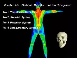
The Skeletal System: The Axial Skeleton. The Skeletal System: The Axial Skeleton. Divisions of the Skeletal System Types of Bones Bone Surface Markings Skull Hyoid Bone Vertebral Column Thorax. Divisions of the Skeletal System. The human skeleton consists of 206 named bones

E N D
The Skeletal System: The Axial Skeleton • Divisions of the Skeletal System • Types of Bones • Bone Surface Markings • Skull • Hyoid Bone • Vertebral Column • Thorax .
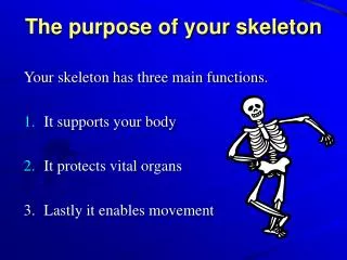
Divisions of the Skeletal System • The human skeleton consists of 206 named bones • Bones of the skeleton are grouped into two principal divisions: • Axial skeleton • Consists of the bones that lie around the longitudinal axisof the human body • Skull bones, auditory ossicles (ear bones), hyoid bone, ribs, sternum (breastbone), and bones of the vertebral column • Appendicular skeleton • Consists of the bones of the upper and lower limbs (extremities), plus the bones forming the girdles that connect the limbs to the axial skeleton .
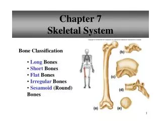
Types of Bones • Bones can be classified into five types based on shape: • Long • Short • Flat • Irregular • Sesamoid
Types of Bones • Long Bones • Greater length than width and are slightly curved for strength • Femur, tibia, fibula, humerus, ulna, radius, phalanges • Short bones • Cube-shaped and are nearly equal in length and width • Carpal, tarsal • Flat bones • Thin and composed of two nearly parallel plates of compact bone tissue enclosing a layer of spongy bone tissue • Cranial, sternum, ribs, scapulae • Irregular bones • Complex shapes and cannot be grouped into any of the previous categories • Vertebrae, hip bones, some facial bones, calcaneus • Sesamoid bones • Protect tendons from excessive wear and tear • Patellae, foot, hand • Sutural bones • Small bones located in sutures of cranial bones
Bone Surface Markings • Bones have characteristic surface markings • Structural features adapted for specific functions • There are two major types of surface markings: • 1) Depressions and openings • Allow the passage of blood vessels and nerves or form joints • 2) Processes • Projections or outgrowths that form joints or serve as attachment points for ligaments and tendons
Skull • Skull (cranium) • Consists of 22 bones • Bones of the skull are grouped into two categories: • Cranial bones • Eight cranial bones form the cranial cavity • Frontal bone, two parietal bones, two temporal bones, the occipital bone, the sphenoid bone, ethmoid bone • Facial bones • Fourteen facial bonesform the face • Two nasal bones, two maxillae, two zygomatic bones, the mandible, two lacrimal bones, two palatine bones, two inferior nasal conchae, vomer
Skull • The cranial and facial bones protect and support special sense organs and the brain • Besides forming the large cranial cavity, the skull also forms several smaller cavities • Nasal cavity • Orbits (eye sockets) • Paranasal sinuses • Small cavities which house organs involved in hearing and equilibrium
Skull • Immovable joints called sutures fuse most of the skull bones together • The skull provides large areas of attachment for muscles that move various parts of the head • Skull and facial bones provide attachment for muscles that produce facial expressions • The facial bones form the framework of the face and provide support for the entrances to the digestive and respiratory systems
Skull (Cranial Bones) • Frontal Bone • Forms the forehead • Parietal Bones • Form the sides and roof of the cranial cavity • Temporal Bones • Form the lateral aspects and floor of the cranium • Occipital Bone • Forms the posterior part and most of the base of the cranium • Sphenoid Bone • Lies at the middle part of the base of the skull • Ethmoid Bone • Located on the midline in the anterior part of the cranial floor medial to the orbits • A major superior supporting structure of the nasal cavity • Contain thin projections called conchae which are lined by mucous membranes • Increased surface area in the nasal cavity helps to humidify inhaled air trapping inhaled particles
Skull (Facial Bones) • Nasal Bones • Form the bridge of the nose • Maxillae • Form the upper jawbone • Form most of the hard palate • Separates the nasal cavity from the oral cavity • Zygomatic Bones • commonly called cheekbones, form the prominences of the cheeks • Lacrimal Bones • Form a part of the medial wall of each orbit • Palatine Bones • Form the posterior portion of the hard palate • Inferior Nasal Conchae • Form a part of the inferior lateral wall of the nasal cavity
Skull (Facial Bones) • Vomer • Forms the inferior portion of the nasal septum • Mandible • Lower jawbone • The largest, strongest facial bone • The only movable skull bone • Nasal Septum • Divides the interior of the nasal cavity into right and left sides • “Broken nose,” in most cases, refers to septal damage rather than the nasal bones themselves • Orbits • Eye socket • Foramina • Openings for blood vessels , nerves , or ligaments of the skull
Skull • Unique Features of the Skull • Sutures, Paranasal sinuses, Fontanels • Sutures • an immovable joint that holds most skull bones together • Paranasal Sinuses • Cavities within cranial and facial bones near the nasal cavity • Secretions produced by the mucous membranes which line the sinuses, drain into the nasal cavity • Serve as resonating chambers that intensify and prolong sounds • Fontanels • Areas of unossified tissue • At birth, unossified tissue spaces, commonly called “soft spots” link the cranial bones • Eventually, they are replaced with bone to become sutures • Provide flexibility to the fetal skull, allowing the skull to change shape as it passes through the birth canal
Hyoid Bone • Does not articulate with any other bone • Supports the tongue, providing attachment sites for some tongue muscles and for muscles of the neck and pharynx • The hyoid bone also helps to keep the larynx (voice box) open at all times
Vertebral Column • Also called the spine, backbone, or spinal column • Functions to: • Protect the spinal cord • Support the head • Serve as a point of attachment for the ribs, pelvic girdle, and muscles • The vertebral column is curved to varying degrees in different locations • Curves increase the column strength • Help maintain balance in the upright position • Absorb shocks during walking, and help protect the vertebrae from fracture
Vertebral Column • Various conditions may exaggerate the normal curves of the vertebral column • Kyphosis • Lordosis • Scoliosis • Composed of a series of bones called vertebrae (Adult=26) • 7 cervical are in the neck region • 12 thoracic are posterior to the thoracic cavity • 5 lumbar support the lower back • 1 sacrum consists of five fused sacral vertebrae • 1 coccyx consists of four fused coccygeal vertebrae
Vertebral Column (Intervertebral Discs) • Found between the bodies of adjacent vertebrae • Functions to: • Form strong joints • Permit various movements of the vertebral column • Absorb vertical shock • Vertebrae typically consist of: • A Body (weight bearing) • A vertebral arch (surrounds the spinal cord) • Several processes (points of attachment for muscles)
Vertebral Column (Regions) • Cervical Region • Cervical vertebrae (C1–C7) • The atlas (C1) is the first cervical vertebra • The axis (C2) is the second cervical vertebra • Thoracic Region • Thoracic vertebrae (T1–T12) • Articulate with the ribs • Lumbar Region • Lumbar vertebrae (L1–L5) • Provide for the attachment of the large back muscles • Sacrum • The sacrum is a triangular bone formed by the union of five sacral vertebrae (S1–S5) • Serves as a strong foundation for the pelvic girdle • Coccyx • The coccyx, like the sacrum, is triangular in shape • It is formed by the fusion of usually four coccygeal vertebrae
Thorax • Thoracic cage is formed by the: • Sternum • Ribs • Costal cartilages • Thoracic vertebrae • Functions to: • Enclose and protect the organs in the thoracic and abdominal cavities • Provide support for the bones of the upper limbs • Play a role in breathing
Thorax • Sternum • “Breastbone” located in the center of the thoracic wall • Consists of the manubrium, body, xiphoid process • Ribs • Twelve pairs of ribsgive structural support to the sides of the thoracic cavity • Costal cartilages • Costal cartilages contribute to the elasticity of the thoracic cage