Download
1 / 27
280 likes | 431 Views
A Class of Membrane Proteins Shaping the Tubular Endoplasmic Reticulum. By: Dorothee van Breevoort Panos Athanasopoulos Nika Strokappe. Main question. How is the shape of the tubular ER formed and maintained? Which proteins are involved What is the mechanism behind it.

E N D
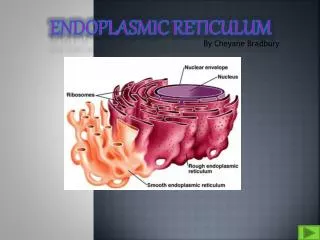
A Class of Membrane Proteins Shaping the Tubular Endoplasmic Reticulum By: Dorothee van Breevoort Panos Athanasopoulos Nika Strokappe
Main question • How is the shape of the tubular ER formed and maintained? • Which proteins are involved • What is the mechanism behind it.
In vitro network formation They used: • Membranes from Xenopus eggs. • Salt wash.
Conclusion Small vesicles (GTP) Large vesicles (High salt wash) Tubuli
Measuring Ca2+ efflux For quantification of the observations
Protein modification effect • MP: Adds PEG • NEM: Adds N-ethyl • MB: Adds biotin • MN: Adds neutravidin • DTT: Has free cysteins
Identification of the target protein • First add biotin, so proteins can be purified. • After adding PEG, some protein disappear. They have a SH group on the surface that is accessible also to PEG
Conclusion • Protein modification prevents the formation of tubuli. • Rtn4a and Rtn4b are the most likely candidates that induce the formation of tubuli.
Conclusion • The modification of proteins by adding biotin correlates with the efflux of Ca2+
Reticulon 4b • Blue: Hydrophobic areas • Green: Area to which antibodies were raised
Conclusion • Antibodies against Rtn4a interfere with the formation of tubuli, antibodies against other ER proteins do not.
Sec61ßER Protein nuclear envelope and reticular network Rtn4a/NogaAPeripheral ER Rtn4c/NogoC Reticular isoform(only reticulon domain)same as Rtn4a/NogaA LocalizationIs Rtn4a/NogoA localized in the ER, more specific in the peripheral ER? Figure 4
Localization (Yeast)Is Rtn4a/NogoA localized in the ER, more specific in the peripheral ER? Figure 4 Conclusion: Reticulons are restricted to the tubular, peripheral ER, consistent with a role in shaping this organelle. • Results • Rtn2/Rtn1 • Absent for NE • Abundant in tubules of peripheral ER
ER structure Figure5 Overexpression Rtna/NogoA ER tubules longer+ less brached Green = Sec61ß (nuclear envelope and reticular network) Red = Rnt4a/NogoA
ER structure Rtn1p • Disruption of peripheral ER • Nuclear envelope intact Together The reticulons have a strong preference to localize to tubular ER When overexpressed reticulons appear to induce tubules Figure 4 Figure 5
MutantsDo yeast cells lacking the reticulons gave altered ER morphology? • Single mutantsNormal • Double mutant :Stress peripheral ERMembrane sheets Conclusion Reticulons are needed for the maintenance of tubular ER under certain stress conditions, but they cannot be the only component required under normal circumstances
Are there additional components involved in shaping the tubular ER?Bindingpartners of Rnt4a/NogoA DP1 Yop1P (Yeast) Blue: Hydrophobic Green: Area to which antibodies were raised Red: Petide identified by mass spec.
localization DP1 DP1 Tubular ER Colocalization Rtn4a/NogoADP1 Figure 6
Is DP1 the missing component for maintaining peripheral ER? ΔreticulonΔyop1 disrupted peripheral ER Some peripheral tubulesminor component Conclusion Rtn1p and Yop1p are the major redundant components required to maintain the tubular ER Figure 6
What is the membrane topology? Two long hydrophobic segments (30-35 ) Rnt4c/NogoCIntroduces single cysteinesW18 andS180 reach by MPEGfirst hydrophobic segments hairspin (second maybe) DP1First hydrophobic segment hairpin ConclusionThe reticulon and DP1 share a rather unusual membrane topology of at least there first hydrophobic segment. Figure 7
SummaryIndication that the reticulons and DP1/Yop1p are ’morphogenic’ proteins that are necessary to form and maintain the tubular ER • Rnt4a/NogoA is required for ER tubule formation in vitro. • Reticulons and DP1/Yop1p localize almost exclusively to the tubular ER,consistent with a role in shaping ER domain • Overexpression of the reticulons leads to long relatively unbranched tubules. • The deletion of the reticulons leads to disruption of the peripheral tubular ER in stress situations and the additional deletion of Yop1p leads to similar ER morphology defects.
Discussion How can salt concentrations affect tubuli formation?
Discussion Is the title of he paper justified by its contents?