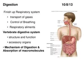
Download
1 / 22
250 likes | 520 Views
Unit II: Transport Breathing Mechanism. Chapter 20 pp 736-759. Pulmonary Ventilation. Breathing – repeating cycle of inspiration and expiration quiet respiration – at rest forced respiration – during exercise

E N D
Unit II: TransportBreathing Mechanism Chapter 20 pp 736-759
Pulmonary Ventilation • Breathing – repeating cycle of inspiration and expiration • quiet respiration – at rest • forced respiration – during exercise • Flow of air in and out of lungs requires a pressure difference between air pressure within lungs and outside body
Pulmonary Ventilation Primary Inspiratory Muscle Accessory Inspiratory Muscles External intercostal muscles Sternocleido- mastoid muscle Accessory Expiratory Muscles Scalene muscles Pectoralis minor muscle Internal intercostal muscles Serratus anterior muscle Transversus thoracis muscle Primary Inspiratory Muscle External oblique muscle Diaphragm Rectus abdominis Internal oblique muscle
Pressure and Flow • Atmospheric pressure drives respiration • 1 atmosphere (atm) = 760 mmHg • Boyle’s Law: • pressure is inversely proportional to volume • for a given amount of gas, as volume , pressure and as volume , pressure • Charles’s Law: • Volume is directly proportional to its absolute temperature • for a given amount of gas, if absolute temperature is doubled, so is the volume
Inspiration • Phrenic nerves stimulate diaphragm to flatten and drop & • intercostal muscles cause ribs to move slightly up and out • volume of thoracic cavity • intrapleuralpressure • visceral pleura clings to parietal pleura • lungs expand with visceral pleura - intrapulmonary pressure • net result: 757 mmHg • Inflation aided by warming of inhaled air • 500 ml of air flows with a quiet breath
Passive Expiration • Expiration achieved by elasticity of thoracic cage • After inspiration, phrenic nerves continue to stimulate diaphragm to produce a braking action to elastic recoil • As volume of thoracic cavity ,intrapulmonary pressure and air is expelled • During quiet breathing: +3mmHg • During forced breathing: +30mmHg
Resistance to Airflow • Diameter of bronchioles • Surface Tension • Thin film of water needed for gas exchange • creates surface tension that would collapse alveoli • Pulmonary surfactant (great alveolar cells) • an agent that disrupts the H+ bonds of water • decreasessurface tension
Alveolar Ventilation • Anatomical dead space • conducting division of airway • Physiological dead space • sum of anatomical dead space + any pathological alveolar dead space • Alveolar ventilation rate • directly relevant to ability to exchange gases
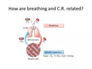
Neural Control of Breathing • Breathing depends on repetitive stimuli from brain • Controlled at 2 levels: • Voluntary control provided by motor cortex • Neurons in medulla oblongata and pons control unconscious breathing
Respiratory Control Centers • Medulla oblongata: • Ventral respiratory group (VRG) • primary generator of respiratory rhythm • Dorsal respiratory group (DRG) • modifies respiratory rhythm • Integrating center
Respiratory Control Centers • Pons: • Pontine respiratory group (PRG) or Pneumotaxic center • regulates shift from inspiration to exhalation • as frequency rises, breathe shorter and shallower
Respiratory Control Centers Forced Breathing Quiet Breathing INHALATION INHALATION (2 seconds) Inspiratory muscles contract, and expiratory muscles relax. Inhalation occurs. Diaphragm and external intercostal muscles contract and inhalation occurs. Increased activity in the DRG stimulates neurons of the VRG that in turn activate the accessory muscles involved in inhalation. The expiratory center of the VRG is inhibited. DRG and inspiratory center of VRG are inhibited. Expiratory center of VRG is active. Neurons in the VRG become inactive. They remain quiet for the next 3 seconds and allow the inspiratory muscles to relax. Activity in the VRG stimulates the inspiratory muscles. After each inhalation, active exhalation occurs as the neurons of the expiratory center of the VRG stimulate the appropriate accessory muscles. Inspiratory muscles relax. Diaphragm and external intercostal muscles relax and passive exhalation occurs. EXHALATION (3 seconds) EXHALATION
Air-Water Interface Example: Soda is put into the can under pressure, and the gas (carbon dioxide) is in solution at equilibrium. • Gases diffuse down their pressure gradients • Henry’s law • amount of gas that dissolves in water is determined by its solubility in water and its partial pressure in air • Temperature of liquid Open the can: internal pressure falls gas molecules come out of solution Volume difference is so great that within a half hour you are left with “flat” soda.
External Respiration Alveolar Gas Exchange Alveolus PO2 = 40 • Exchange across the water film covering alveolar epithelium and respiratory membrane • Factors affecting gas exchange: • Pressure gradients of gases PCO2 = 45 Respiratory membrane Pulmonary circuit Systemic circuit PO2 = 100 PCO2 = 40 PO2 = 100 mm Hg Pulmonary capillary PCO2 = 40 mm Hg Internal Respiration Interstitial fluid Systemic circuit PO2 = 95 PCO2 = 40 PO2 = 40 PCO2 = 45 PO2 = 40 PCO2 = 45 Systemic capillary
Other Factors Affecting Gas Exchange • Gas solubility • CO2 20 times as soluble as O2 • Membrane thickness - only 0.5 m thick • Membrane surface area - 100 ml blood in alveolar capillaries, spread over 70 m2 • Ventilation-perfusion coupling - areas of good ventilation need good perfusion (vasodilation)
Oxygen Transport • Concentration in arterial blood • 98.5% bound to hemoglobin • 1.5% dissolved • Binding to hemoglobin • each heme group of 4 globin chains may bind O2 • oxyhemoglobin (HbO2 ) • deoxyhemoglobin (HHb) Protein subunits Iron ion Heme unit
Oxyhemoglobin Dissociation Curve Steep slope = a large change in amount of oxygen associated with Hb. Blood entering the systemic circuit has a PO2 of 95 mm Hg. Blood leaving peripheral tissues has an average PO2 of 40 mm Hg. Oxyhemoglobin (% saturation) The PO2 in active muscle tissue may drop to 15–20 mm Hg. PO2 (mm Hg)
Oxyhemoglobin Dissociation Curve Bohr Effect pH increase = curve shifts to the left Hemoglobin releases less oxygen. 7.6 7.4 7.2 pH decrease = curve shifts to the right Hemoglobin releases more oxygen. Oxyhemoglobin (% saturation) PO2 (mm Hg)
CO2 diffuses into the bloodstream. Carbon Dioxide Transport ~7% remains in solution. 93% diffuses into RBCs. ~70% is converted to carbonic acid by carbonic anhydrase ~23% CO2 is carried as Carbaminohemoglobin (HbCO2) H2CO3 H+ + HCO3– Hydrogen ions bind to hemoglobin acting as pH buffers. Bicarbonate ions move into the surrounding plasma in exchange for extracellular chloride ions (Cl–). Chloride shift
Summary of Transport O2 pickup O2 delivery Pulmonary capillary Systemic capillary Plasma Red blood cell Red blood cell Cells in peripheral tissues Alveolar air space Chloride shift Alveolar air space Pulmonary capillary Systemic capillary CO2 delivery CO2 pickup