Download
1 / 40
510 likes | 988 Views
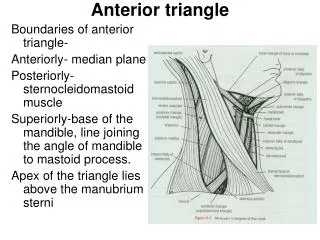
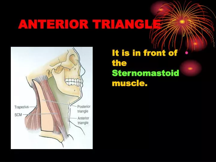
ANTERIOR TRIANGLE. It is in front of the Sternomastoid muscle. BOUNDARIES. Anteriorly : midline of the neck. Posteriorly : anterior border of sternomastoid. Superiorly : lower margin of the body of the mandible. BOUNDARIES. Roof :

E N D
ANTERIOR TRIANGLE • It is in front of the Sternomastoid muscle.
BOUNDARIES • Anteriorly: midline of the neck. • Posteriorly: anterior border of sternomastoid. • Superiorly: lower margin of the body of the mandible.
BOUNDARIES • Roof: • skin, superficial fascia (containing platysma) and theInvesting layerof deep cervical fascia.
SUBDIVISIONS • The anterior triangle is subdivided by : • The Anterior and Posterior bellies of the Digastric and • The Superior belly of Omohyoid into (4) smaller triangles.
SUBMENTAL TRIANGLE • It lies below the chin. • Boundaries : • Anteriorly: midline of the neck. • Laterally: Anterior belly of Digastric.
BOUNDARIES • Inferiorly : body of the hyoid. • Floor: Mylohyoid muscle.
CONTENTS • Submental lymph nodes. • They receive lymph from the tip of the tongue.
MYLOHYOID MUSCLE • It is a flat triangular sheet that supports the floor of the mouth and tongue (forms the main part of the floor of the mouth). • Origin : • Mylohyoid line of the mandible.
MYLOHYOID • Insertion: • The anterior fibers: Into a Fibrous Raphe. • The posterior fibers: Into the body of hyoid bone.
MYLOHYOID • Nerve supply : • Nerve to mylohyoid.
MYLOHYOID • Action : • If the mandible is fixed:Elevates the floor of the mouth as in deglutition. • If the hyoid bone is fixed:Depresses the mandible and opens the mouth.
DIGASTRIC TRIANGLE • It lies below the body of the mandible. • Boundaries: • Anteriorly: • Anterior belly of Digastric. • Posteriorly: • Posterior belly of digastric & Stylohyoid.
DIGASTRIC TRIANGLE • Superiorly: • Lower border of the body of the mandible. • Floor: • Mylohyoid. Hyoglossus.
CONTENTS • A. Anterior part : • (1) Submandibularsalivarygland. • (2) Facial artery (deep to gland). • (3) Facial vein • (4) Submandibular lymphnodes. • The vein and lymph nodes are superficial to the gland.
CONTENTS • (5) Hypoglossal nerve. • (6) Nerve and vessels to mylohyoid.
CONTENTS • B. posterior part: • 1. Carotid sheath and its contents. • 2.parotid gland (Lower part)
DIGASTRIC MUSCLE • It has two bellies: • Anterior and Posterior. • Posterior belly : • Arises from the mastoid process. • Inserted into the intermediate tendon. • Anterior belly attached to the lower border of body of mandible.
DIGASTRIC MUSCLE • Insertion : to the • Intermediate Tendon. • It pierces the insertion of stylohyoid. • It binds to the hyoid bone by a loop of deep fascia.
NERVE SUPPLY • Posterior belly: Facial Nerve • Anterior belly: • The nerve to Mylohyoid.
ACTION Depression of the mandible. OR • Elevation of the hyoid bone.
STYLOHYOID MUSCLE • Origin: • The styloid process. • It runs along the upper border of the posterior belly of digastric. • Insertion: • Hyoid bone (between body and greater horn).
STYLOHYOID MUSCLE • Nerve supply : • Facial nerve. • Action : • Elevation of hyoid bone.
CAROTID TRIANGLE • It is behind the hyoid bone. • Boundaries : • Superiorly: • Posterior belly of Digastric. • Inferiorly: • Superior belly of Omohyoid.
BOUNDARIES • Posteriorly: • Anterior border of sternomastoid. • Floor: • Thyrohyoid.Hyoglossus. • Middle & Inferior constrictors of the pharynx.
CONTENTS • (1) Carotid sheath • (2) Hypoglossal nerve and its descending branch. • (3) Acessory nerve.
CONTENTS • (4) Internal and External laryngeal nerves. • (5) Deep cervicallymph nodes.
CAROTID SHEATH • It is a condensation of deep cervical fascia. • It is attached to the base of the skull superiorly and fuses with the pericardium inferiorly.
CONTENTS • 1. Common and internal carotid arteries. • 2. Internal jugular vein. • 3. Vagus nerve. • 4. Deep cervical lymph nodes.They form a chain along the internal jugular vein.
CAROTID SHEATH • It is crossed by • Glossopharyngeal. • Hypoglossal. • Spinal part of acessory.
MUSCULARTRIANGLE • It lies below the hyoid bone. • Boundaries: • Anteriorly : • Midline of the neck. • Superiorly: • Superior belly of Omohyoid.
MUSCULAR TRIANGLE • Inferiorly: • Anterior border of Sternomastoid • Floor: • Sternohyoid.Sternothyroid.
FLOOR • Beneath the floor lie: • Thyroid gland. • Larynx. • Trachea. • Esophagus.
ORIGIN • Sternohyoid & Sternothyroid : • Posterior surface of the manubrium. • Thyrohyoid: • Oblique line of thyroid cartilage.
ORIGIN • Omohyoid : • Inferior belly : • Suprascapular ligament and Suprascapular notch. • Superior belly: • Intermediate tendon.
INSERTION • Sternohyoid & Sternothyroid : • Hyoid bone (lower border). • Oblique line of thyroid cartilage. • Thyrohyoid: • Hyoid bone (lower border).
INSERTION • Omohyoid • Hyoid bone (lower border).
NERVE SUPPLY • Ansa Cervicalis (C1,2&3) • EXCEPT • Thyrohyoid: • (C1) through the hypoglossal nerve.
ACTION • (1) stabilization of the hyoid bone to make a base for the movements of the tongue. • (2) Assistance in the movements of the larynx in swallowing.