Download
1 / 62
650 likes | 1.04k Views
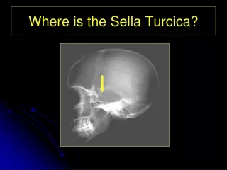
Sella Turcica and Parasellar Region. F.Niaghi , MD Radiologist. Anatomic Approach to Differential Diagnosis. In order to analyze a sellar or parasellar mass on MRI we use the following anatomic approach: First identify the pituitary gland and sella turcica .

E N D
Sella Turcica and Parasellar Region F.Niaghi, MD Radiologist
Anatomic Approach to Differential Diagnosis In order to analyze a sellar or parasellar mass on MRI we use the following anatomic approach: • First identify the pituitary gland and sellaturcica. • Then determine the epicenter of the lesion and whether it is in the sella or above, below or lateral to the sella. • If it is in the sella, determine whether or not the sella is enlarged. • Once the location of the mass is clear, analyze the signal intensity patterns: is the lesion cystic or solid? • Does it contain any abnormal vessels? • Are there any calcifications? And so on. • Finally establish a Differential Diagnosis.
Pituitary gland • On a coronal section through the brain the reference structure is the pituitary gland which lies in the sellaturcica. • It is usually larger in females than in males - in females the superior border tends to be convex, whereas in males it is usually concave. • The most common abnormalities that arise in the pituitary gland are pituitary adenoma, Rathke's cleft cyst and craniopharyngioma.
Pituitary stalk • The next structure to identify is the pituitary stalk. • This is a vertically oriented structure which connects the pituitary gland to the brain. • It is thinner at the bottom and thicker at the top. • Embryologically, it is also derived from Rathke's cleft epithelium and therefore the pathologies, which can arise in the pituitary gland can also arise in the stalk. • There are a few unusual things to be considered in children, such as germinomas and eosinophilic granulomas. • In adults metastases and occasionally lymphoma can arise in the pituitary stalk
Optic chiasm • Another major structure in the suprasellar cistern is the optic chiasm. • It is an extension of the brain and looks like the number 8 lying on its side. • It is glial tissue - therefore the most common tumors to originate here are gliomas. • Another frequent pathology in this region is demyelinating disease - particularly multiple sclerosis. This can also be associated with some swelling of the optic chiasm.
Hypothalamus • Further cephalad lies the base of the brain, which at this location is the hypothalamus. • Anatomically the hypothalamus forms the lateral walls and floor of the third ventricle. • The most common pathologies to arise here are gliomas - in children hamartomas, germinomas and eosinophilic granuloma.
Carotid artery • A very important structure in this area is the internal carotid artery. • It runs a complex anatomic course as it passes through the skull base shaped like an S on lateral views. • It passes through the cavernous sinus. • The segment cranial to this is known as the supracavernous segment. • This bifurcates into the anterior cerebral artery, which passes cranially to the optic chiasm, and the middle cerebral artery, which runs laterally. • Aneurysms and ectasias are pathologies that can arise here. • One must also be aware of congenital variations in the course of the internal carotid Sometimes it is very medially positioned and can actually lie in the midline.
Cavernous sinus • The cavernous sinus is a paired complex of venous channels. • In the lateral wall of the sinus run nerve III (oculomotorius), IV (trochlearis), V1 and V2 (trigeminus). • The sixth cranial nerve (abducens) runs more medially and is located caudal to the carotid artery. • The most common pathologies occurring in the cavernous sinus include schwannomas arising from the cranial nerves and inflammation, which can lead to thrombosis. • This is known as cavernous sinus thrombophlebitis. • Carotid-cavernous fistulas are fistulous communications between the carotid artery and the veins of the cavernous sinus.
Meninges • The meninges cover the cavernous sinus. • They are thicker laterally and superiorly than medially and inferiorly. • The most common tumor to arise from the meninges is of course the meningioma. • Dural metastasis is the second most common tumor to arise here. • Also inflammatory pathologies occur in the basal meninges - the most common infection being tuberculous meningitis. • Of the non-infectious inflammatory pathologies sarcoidosis is the commonest.
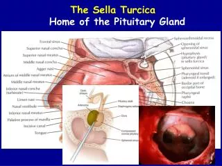
Sphenoid sinus • Inferior to the pituitary gland lies the sphenoid sinus. • This structure contains air and is lined by mucosa and bone. • Posterior to the sphenoid sinus lies the clivus. • Pathology that arises in this area includes carcinomas arising from the mucosa of the sphenoid sinus - squamous cell carcinoma and adenoid cystic carcinoma are the most common. • Chordomasarise in the clivus and chondrosarcomas and osteosarcomas also occur in this area. • Metastases can occur anywhere. • Bacterial or fungal inflammatory processes in the sphenoid sinus can spread intracranially via the cavernous sinus.
Pituitary Microadenoma • By definition, pituitary microadenomas are less than 10 mm in diameter and are located in the pituitary gland. • The differential diagnosis: pituitary microadenoma or Rathke's cleft cyst (the two can be indistinguishable). • The sensitivity of an unenhanced MRI scan for detecting pituitary microadenomas is about 70%. • It is not always necessary to give intravenous contrast for detecting pituitary microadenomas as patients with a negative scan generally receive the same symptomatic treatment as patients with a microadenoma. • The purpose of the scan is to rule out any large lesions. • In possible surgical candidates (for example patients with failed medical therapy or pituitary disease not amenable to medical therapy such as Cushing's disease) it is necessary to give contrast to localize the lesion as accurately as possible.
Pituitary Microadenoma • On an unenhanced scan, approximately 70% of all pituitary microadenomas can be detected. • If you give gadolinium, you can reduce the false-negative rate from 30% to 15%. • As mentioned earlier, this usually does not affect patient management.
Pituitary Macroadenoma • By definition, pituitary macroadenomas are adenomas over 10mm in size. • They tend to be soft, solid lesions, often with areas of necrosis or hemorrhage as they get bigger. • As they grow, they first expand the sellaturcica and then grow upwards. • Because they are soft tumors, they usually indent at the diaphragmasellae, giving them a 'snowman' configuration. • This is one feature that can help distinguish between a pituitary macroadenoma and a meningioma. • Another feature which can help differentiate them is enlargement of the sellaturcica - this generally only occurs with pituitary macroadenomas that originate in the sella.
Cavernous sinus invasion • It is not always possible to tell if there is cavernous sinus invasion, but there are three signs to look out for: • -Is there more than 50% encirclement of the carotid artery? • Note: meningiomas tend to constrict the carotid artery, macroadenomas do not. • -Is there lateral displacement of the lateral wall of the cavernous sinus compared to the opposite side? • -Is there an increased amount of tissue interposed between the carotid artery and the lateral wall of the cavernous sinus?
Rathke's cleft cyst • Rathke's cleft cyst is the second of three pathologies derived from Rathke's cleft epithelium. • The cyst is fluid-filled and has very thin walls with a thickness of only one or two cell layers. • These walls can contain cells which secrete fluid, allowing the cyst to grow and compress adjacent structures. • Rathke'scleft cysts can occur either in or above the sellaturcica.
Craniopharyngioma • Craniopharyngioma is the third of the three pathologies derived from Rathke's cleft epithelium. • Technically these are benign tumors, but unlike Rathke's cleft cysts, they have thick walls and are locally invasive. • Macroscopically, it is a complex mass with multiple nodules at the base of the brain, sinuating along the fissures. • Often, it can not be completely resected.
Craniopharyngioma • In over 50% of cases craniopharyngiomas have a pathognomonic appearance. • A compressed pituitary gland can be identified. • There is a large intrasellar and suprasellar mass with cystic and enhancing components as well as calcifications. • These findings in a child are virtually pathognomonic for craniopharyngioma (perhaps with only a dermoid in the differential diagnosis).
Meningioma • The most common intracranial tumor in adults is the meningioma with 20% of occurring at the skull base. • Meningiomasare almost always solid lesions, sometimes with a cyst on the edge. • They can lift up the arachnoid a little bit and enhance uniformly as a general rule.
On the top-left unenhanced and enhanced CT-images, the main differential diagnosis of the enhancing mass would include meningioma, pituitary adenoma and an aneurysm. The post-constrast MR-image on the top-right rules out an aneurysm as a possible diagnosis (no flow void), but on axial images a pituitary adenoma and meningioma are still difficult to differentiate
Notice the spread of the lesion along the meninges.The epicentre of the lesion is above the sella.
Note the compresses pituitary gland There is no evidence of cystic change or hemorrhage
Thrombosed aneurysm On the left the T1-weighted image of a thrombosed aneurysm with high signal intensity on the unenhanced scan. It originates in the intracavernous segment of the right internal carotid artery. On the right the T2-weighted images: the thrombosed aneurysm has a dark rim.
Hamartoma • Hamartomas are masses of dysplastic tissue found almost exclusively in young children. • One of the most common locations is the floor of the third ventricle. • They are benign lesions, but patients do succumb to them because of the bad location.