Download
1 / 21
220 likes | 375 Views
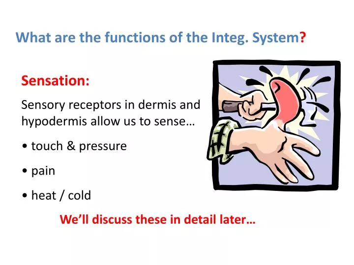
What are the functions of the Integ. System ?. Sensation:. Sensory receptors in dermis and hypodermis allow us to sense… touch & pressure pain heat / cold. We’ll discuss these in detail later…. What are the functions of the Integ. System ?. Excretion:.

E N D
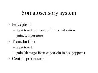
What are the functions of the Integ. System? Sensation: • Sensory receptors in dermis and hypodermis allow us to sense… • touch & pressure • pain • heat / cold We’ll discuss these in detail later…
What are the functions of the Integ. System? Excretion: Small amounts of nitrogenous waste products (NH3, Urea etc.) are lost through sweating. Insignificant compared to excretory system!!!
Hypodermis Technically NOT part of the skin, varies in thickness depending in part on the amount of adipose present What are the main layers of the Integ. System? 2 (3) main layers:EpidermisThin layer of stratified squamous epithelium DermisThicker, tough layer of dense irregular connective tissue and other components
Take 2!!! What layer(s) of the epidermis allow this baby to have hope of “normal” face again? Discuss with your neighbor and predict an answer.
What are the Dermis Details? • Contains all the “goodies” • 2 layers Reticular layerPapillary layer Dermis is primarily what type of tissue? Consists of fibroblasts and abundant protein fibers (matrix), adipose cells and macrophages What types of fibers? Reticular Elastic Collagen
How, where and why do we have “fingerprints”? Epidermis of some skin follows contours of parallel papillary ridges. Is this in thin or thick skin?
Is the skin equally strong in all directions? Tension (cleavage) lines Striae (stretch marks) As the skin is stretched the dermis “ruptures” and forms linear scars (striae). Let’s predict the orientation of tension lines based on the direction of striae!
What are the Hypodermis Details? • Fibroblasts, adipose cells (1/2 of body fat) and macrophages • Primarily loose connective tissue • Also called subcutaneous tissue
What type of glands are in the skin? • Sweat glands… • Merocrine sweat glands • Apocrine sweat glands Abundant & widespread, mostly H2O, ducts to surface of skin, myoepithelial cells. Less abundant, found in axillary, anal, pubic regions (& faces of men), > conc. Of fatty acids, bacteria breakdown cause rancid smell, ducts to hair follicles.
What type of glands are in the skin? Sebaceous glands… Secrete sebum (oily substance) often into hair follicle (holocrine glands see pg. 113). Other glands… Ceruminous Mammary
What’s all the fuzz about? Growth from Matrix!
How do you explain Deed’s foot? What would cause that color? Is the lack of sensation a symptom you would attribute to that color? Why or why not? What parts of the scene are “fakey”?
What is the reality? Developing gangrene risks: Chronic lack of sufficient blood flow! Conditions causing this: Diabetes Atherosclerosis Thrombosis - a clot in a blood vessel, also related to atherosclerosis Extreme cold injury (frostbite). http://www.reversegangrene.com/A.htm
Eschar Why are burns some of the most painful injuries? Burns are ranked based on the severity and the depth of tissue destruction. 1st degree:Epithelium damaged ~red & painful, NO blisters 2nd degree:Epithelium & some dermis damaged ~red, white or tan, very painful, BLISTERS present 3rd degree (full thickness)All layers damaged ~black, NO pain, NO blisters
Is all skin cancer equally dangerous? Types are identified based on the type of cells that mutate:Basal cell carcinoma Least dangerous, start as “shiny” bump, center becomes concave and ulcerus Squamous cell carcinoma Raised, red, “scaly” & ulcerus, can metastasize Melanoma Dark, flat, irregular, most deadly
LEARNING OUTCOMES 11.4 Integumentary System *Identify the two main regions of skin, and how these are distinguished from the subcutaneous layer. *Describe the makeup and function of the accessory structures of human skin. *List some common disorders of human skin, and how these may be treated.